Copyright
©2014 Baishideng Publishing Group Inc.
World J Gastroenterol. Sep 28, 2014; 20(36): 13200-13204
Published online Sep 28, 2014. doi: 10.3748/wjg.v20.i36.13200
Published online Sep 28, 2014. doi: 10.3748/wjg.v20.i36.13200
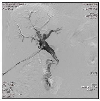
Figure 1 Lower segment stricture of the common bile duct and choledochoduodenal fistula were detected by T-tube cholangiography 6 mo after operation.
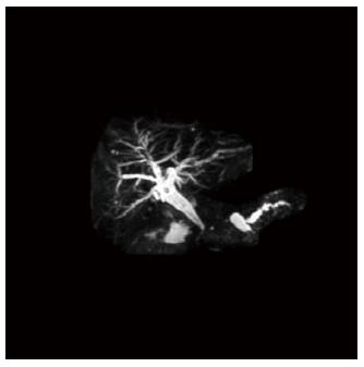
Figure 2 Magnetic resonance cholangiopancreatography showed discontinuity of the pancreatic duct 6 mo after operation.
The duct in the pancreatic head and neck was not detected and pancreatic leakage from the proximal duct of the pancreatic body and dilation of the distal duct of the pancreatic body and tail were not observed.
Figure 4 Fifteen centimeters of the jejunum to the Treitz ligament was transected with side closures.
A: A retrocolic end-to-side pancreaticojejunostomy was then performed using a distal jejunal limb. At 20 cm distal to the pancreaticojejunostomy, the jejunum was interrupted with side closures. Twenty centimeters of the jejunum to the pancreaticojejunostomy was transected with side closures. An antecolic end-to-side hepaticojejunostomy was then performed using a distal jejunal limb. Then the first enteroenterostomy was performed at 20 cm to the hepaticojejunostomy using a distal jejunal limb to the pancreaticojejunostomy and the second enteroenterostomy was performed at 20 cm to the first enteroenterostomy using a distal jejunal limb to the Treitz ligament (curved arrow in A: jejunal limb used for hepaticojejunostomy; arrow in A: first enteroenterostomy; triangular arrow in A: second enteroenterostomy); B: Schematic diagram of the pancreaticojejunostomy, hepaticojejunostomy and double Roux-en-Y digestive tract reconstruction.
- Citation: Jia CK, Lu XF, Yang QZ, Weng J, Chen YK, Fu Y. Pancreaticojejunostomy, hepaticojejunostomy and double Roux-en-Y digestive tract reconstruction for benign pancreatic diseases. World J Gastroenterol 2014; 20(36): 13200-13204
- URL: https://www.wjgnet.com/1007-9327/full/v20/i36/13200.htm
- DOI: https://dx.doi.org/10.3748/wjg.v20.i36.13200


