Copyright
©2014 Baishideng Publishing Group Inc.
World J Gastroenterol. Sep 28, 2014; 20(36): 13191-13194
Published online Sep 28, 2014. doi: 10.3748/wjg.v20.i36.13191
Published online Sep 28, 2014. doi: 10.3748/wjg.v20.i36.13191
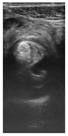
Figure 1 Axial ultrasound image showing small bowel intussusception.
The echogenic mesenteric fat is seen trapped between the intussusceptum and the intussuscipiens.
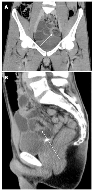
Figure 2 Corresponding coronal computed tomography (A) and sagittal computed tomography (B) images.
Computed tomography images obtained after administration of intravenous contrast material show the dilated fluid-filled bowel loops (arrows) as well as the intussusceptum and the intussuscipiens in the left lower quadrant, with trapped mesenteric fat.
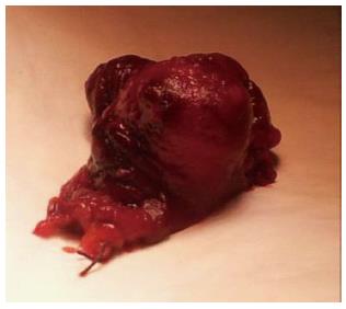
Figure 3 Post-operative specimen of ileum segment with granuloma.
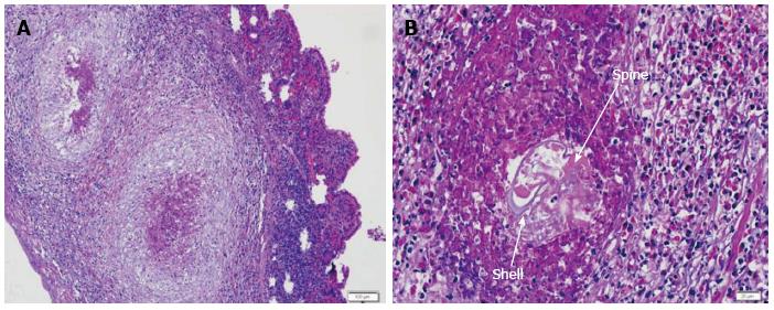
Figure 4 Schistosoma species.
A: Bowel wall with granuloma formation (HE stain); B: A 75 μm-wide ovoid structure with a thin and basophilic shell (arrow) and presenting a spine (HE stain).
- Citation: Pinto JP, Cordeiro A, Ferreira AM, Antunes C, Botelho P, Rodrigues AJ, Leão P. Ileal intussusception due to a parasite egg: A case report. World J Gastroenterol 2014; 20(36): 13191-13194
- URL: https://www.wjgnet.com/1007-9327/full/v20/i36/13191.htm
- DOI: https://dx.doi.org/10.3748/wjg.v20.i36.13191


