Copyright
©2014 Baishideng Publishing Group Inc.
World J Gastroenterol. Sep 28, 2014; 20(36): 13105-13118
Published online Sep 28, 2014. doi: 10.3748/wjg.v20.i36.13105
Published online Sep 28, 2014. doi: 10.3748/wjg.v20.i36.13105
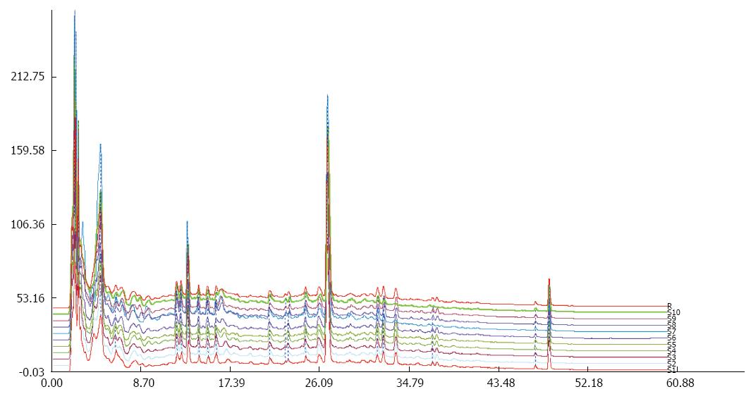
Figure 1 High-performance liquid chromatography-DAD of the samples.
The samples were randomly tested ten times, and the absorbance was read at 280 nm.
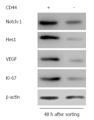
Figure 2 Results of Western blot 48 h after FACS sorting, indicating significant differences in Notch-1, Hes1, VEGF and Ki-67 expression between CD44+ and CD44- cells.
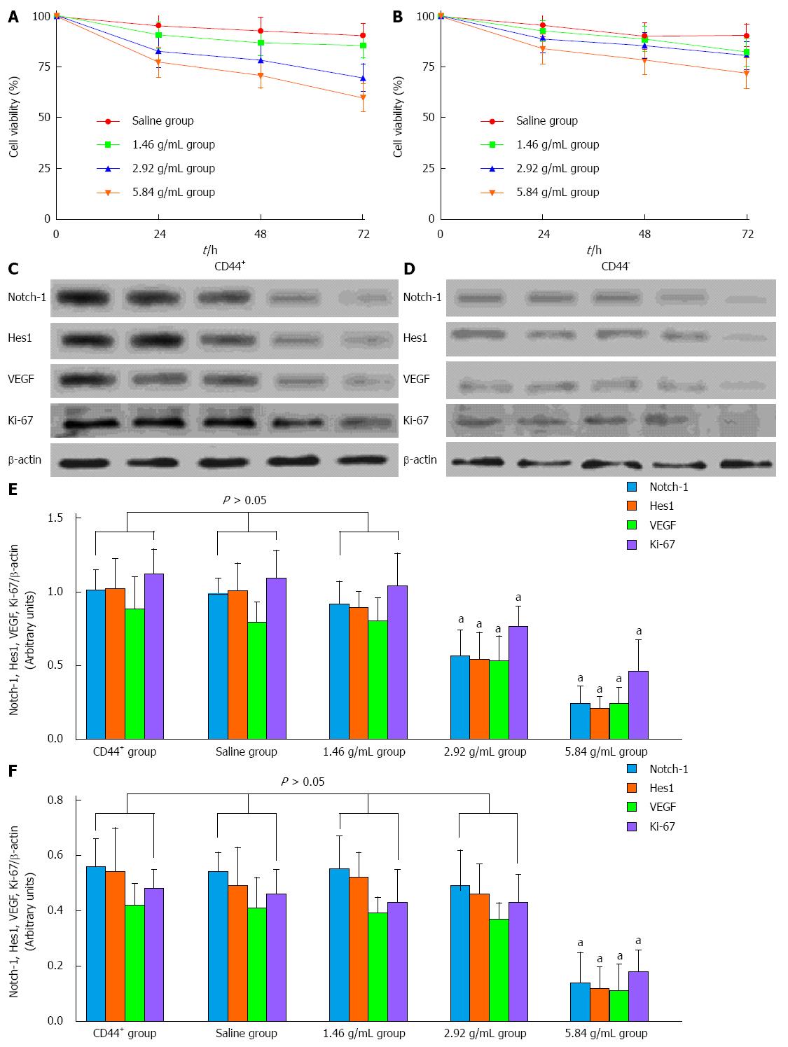
Figure 3 Xiaotan Sanjie decoction inhibits cell viability in a dose-dependent manner.
A, B: The effect of Xiaotan Sanjie (XTSJ) decoction on the viability of CD44+ and CD44- cells. The cells were treated with XTSJ decoction for 24, 48 or 72 h. Cell viability was determined by MTT assay. Dots: Mean of six independent experiments; bars: SD; C, D: Results of the Western blot for the expression of Notch-1, Hes1, VEGF, and Ki-67 in the cells incubated with XTSJ decoction for 48 h; E, F: The statistical analysis of the expression of Notch-1, Hes1, VEGF and Ki-67. The densitometry analysis results of the displayed bands were normalized to β-actin; the data are expressed as arbitrary units (one-way ANOVA, followed by the Bonferroni test). aP < 0.05 vs CD44+ group.
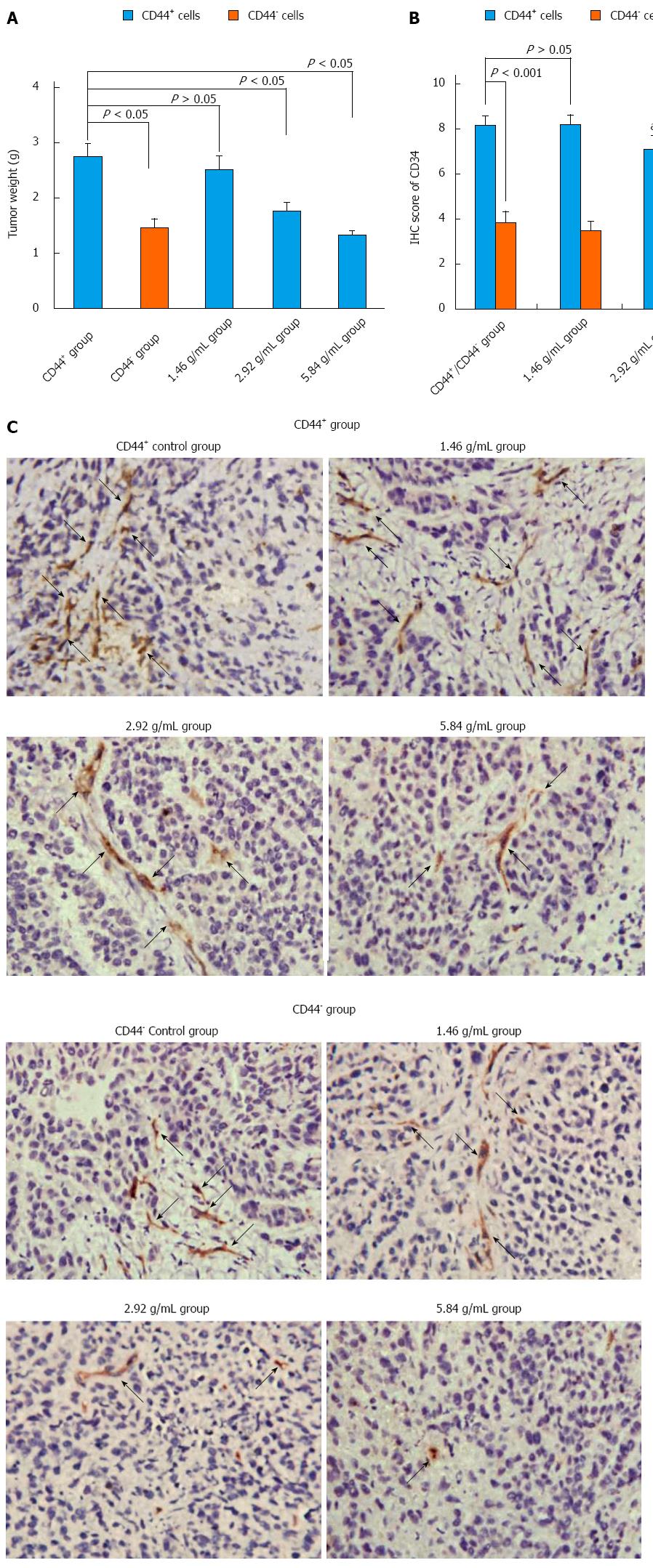
Figure 4 Xiaotan Sanjie decoction inhibits tumor weight and angiogenesis in a dose-dependent manner.
A: Tumor weight after eight consecutive weeks of treatment (n = 6 in each group). Control CD44+ group had larger tumors than control CD44- group (Student’s t-test). Treatment with Xiaotan Sanjie (XTSJ) decoction inhibited tumor growth in a dose-dependent manner in gastric cancer stem-like cell transplanted mice (one-way ANOVA, followed by the Bonferroni test); B: Comparison of the microvessel density (MVD) in different groups. The control CD44+ mice showed higher MVD than the control CD44- mice (n = 6, P < 0.05), and XTSJ decoction reduced the MVD in a dose-dependent manner. Statistically significant differences in MVD could be detected in the medium- and high-dose CD44+ groups (Nonparametric tests); C: Immunohistochemical (IHC) results of the MVD. The MVD was defined as a discrete CD34-positive endothelial cell aggregate, with or without definable lumina. (original magnification × 400, positive areas are indicated by black arrows). aP < 0.05 vs CD44+ group; cP < 0.05 vs CD44- group.
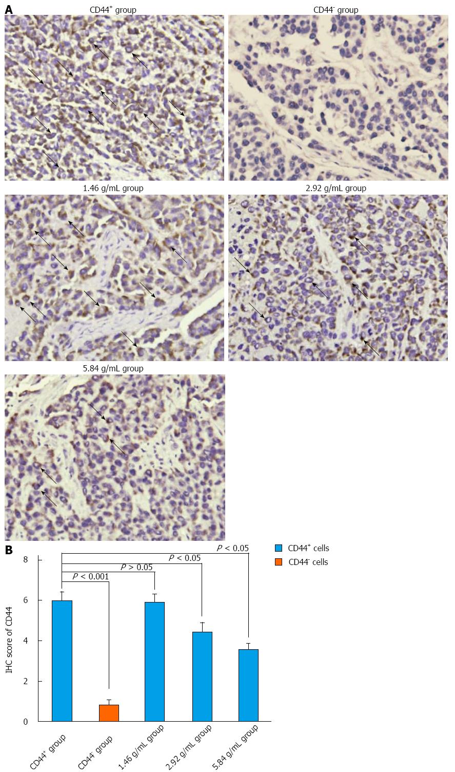
Figure 5 Xiaotan Sanjie decoction inhibits CD44 expression in a dose-dependent manner.
A: Immunohistochemical (IHC) staining for CD44. CD44 was highly expressed in the cancer cell membrane (original magnification × 400, positive areas are indicated by black arrows); B: The comparison of CD44 in different groups. CD44 was highly expressed in the control CD44+ group compared with the rest of the groups and was rarely detected in the control CD44- group. The results also showed that not all of the cells in the CD44+ group were CD44 positive. Xiaotan Sanjie decoction inhibited the expression of CD44 in a dose-dependent manner. Statistically significant differences in the expression of CD44 were also detected in the medium- and high-dose groups in CD44+ cells (nonparametric tests).
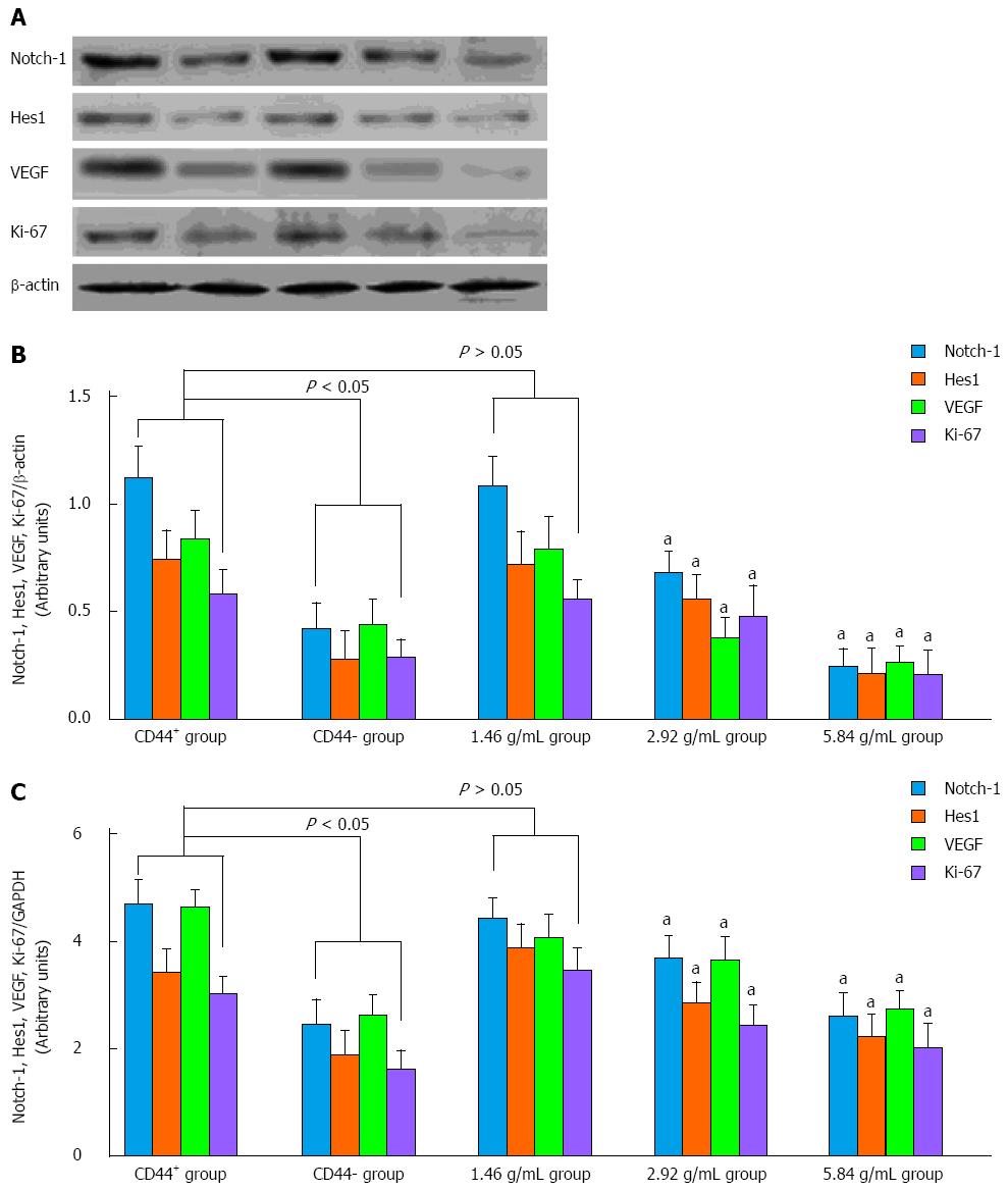
Figure 6 Western blot and real-time quantitative polymerase chain reaction analysis of Notch-1, Hes1, VEGF and Ki-67 expression in vivo.
A, B: Western blot revealed that Notch-1 and Hes1 were highly expressed in the control CD44+ group compared with the control CD44- group. Xiaotan Sanjie decoction inhibited the expression of Notch-1 and Hes1 in a dose-dependent manner, and significant differences could be detected in the medium- and high-dose groups (P < 0.05); C: The variation in Notch-1, Hes1, VEGF and Ki-67 expression in the experimental groups was confirmed by quantitative polymerase chain reaction (one-way ANOVA, followed by the Bonferroni test). aP < 0.05 vs CD44+ group.
- Citation: Yan B, Liu L, Zhao Y, Xiu LJ, Sun DZ, Liu X, Lu Y, Shi J, Zhang YC, Li YJ, Wang XW, Zhou YQ, Feng SH, Lv C, Wei PK, Qin ZF. Xiaotan Sanjie decoction attenuates tumor angiogenesis by manipulating Notch-1-regulated proliferation of gastric cancer stem-like cells. World J Gastroenterol 2014; 20(36): 13105-13118
- URL: https://www.wjgnet.com/1007-9327/full/v20/i36/13105.htm
- DOI: https://dx.doi.org/10.3748/wjg.v20.i36.13105


