Copyright
©2014 Baishideng Publishing Group Inc.
World J Gastroenterol. Sep 28, 2014; 20(36): 13035-13043
Published online Sep 28, 2014. doi: 10.3748/wjg.v20.i36.13035
Published online Sep 28, 2014. doi: 10.3748/wjg.v20.i36.13035
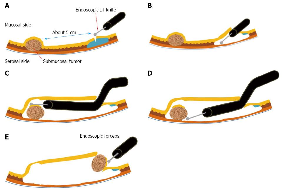
Figure 1 Endoscopic submucosal tunnel dissection.
A: After the mucosa is lifted by submucosal injection, a 2 cm mucosal incision is made approximately 5 cm proximal to the submucosal tumor (SMT); B: A submucosal tunnel is created using endoscopic submucosal dissection; C: The submucosal dissection is continued until the SMT is visible by endoscopy; D: Dissection is performed along the margin of the SMT with an endoscopic knife; E: The dissected SMT is removed through the mucosal defect.
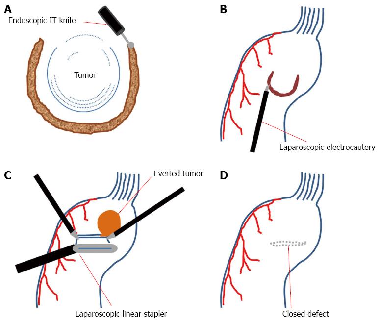
Figure 2 Laparoscopic endoscopic cooperative surgery.
A: Three-fourths of the circumference is cut in the endoscopic side; B: Laparoscopic seromuscular dissection is performed along the submucosal dissection line; C: With the tumor and the non-resected portion are lifted, the defect is closed with laparoscopic linear staplers; D: The direction of stapling should be perpendicular to the longitudinal axis of the stomach.
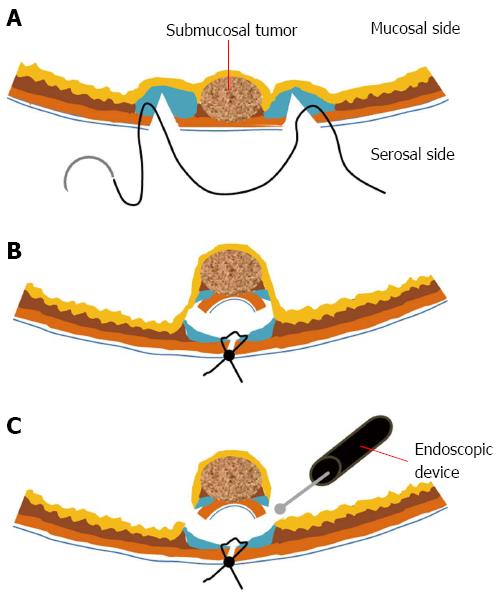
Figure 3 Non-exposed endoscopic wall-inversion surgery.
A: Laparoscopic seromuscular dissection is done after endoscopic submucosal injection. Then, laparoscopic seromuscular suture is performed around the dissected portion; B: Subsequently, the dissected portion is invaginated to the luminal side; C: The mucosal layer is cut with the endoscopic device.
- Citation: Lee CM, Kim HH. Minimally invasive surgery for submucosal (subepithelial) tumors of the stomach. World J Gastroenterol 2014; 20(36): 13035-13043
- URL: https://www.wjgnet.com/1007-9327/full/v20/i36/13035.htm
- DOI: https://dx.doi.org/10.3748/wjg.v20.i36.13035


