Copyright
©2014 Baishideng Publishing Group Inc.
World J Gastroenterol. Aug 28, 2014; 20(32): 11451-11455
Published online Aug 28, 2014. doi: 10.3748/wjg.v20.i32.11451
Published online Aug 28, 2014. doi: 10.3748/wjg.v20.i32.11451
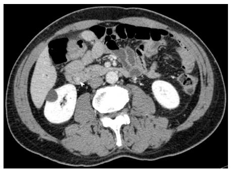
Figure 1 Abdominal computed tomography angiography image of the common bile duct.
Thickening of the wall of the distal common bile duct was observed (arrowhead).
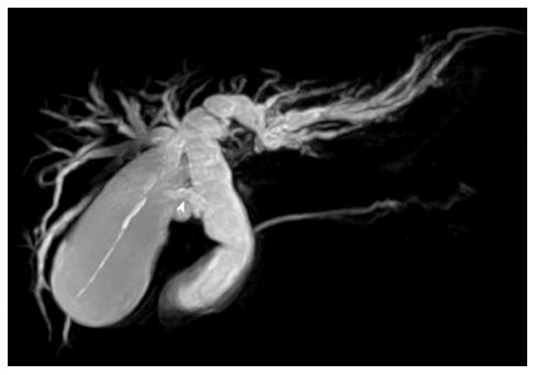
Figure 2 Magnetic resonance cholangiopancreatography.
The common and intrahepatic bile ducts were dilated. It was impossible to detect the accessory bile duct preoperatively (arrowhead).
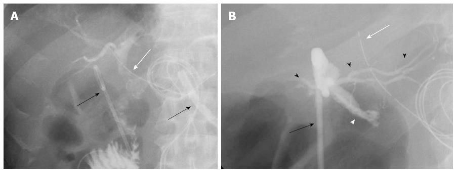
Figure 3 Tubography through the biliary drainage tube (white arrow) and the abdominal drainage tube (black arrow) after the first operation.
A: The radiological image indicated the intrahepatic bile duct, common bile duct, and jejunum. Leakage of the contrast media from the end to side biliojejunostomy was not observed. Two abdominal drainage tubes (black arrows) were inserted into the abdominal cavity; B: Postoperative tubography through the abdominal drainage tube (black arrow) was performed, which produced the images of the injured site of the probable caudate bile duct (white arrowhead) and the other intrahepatic bile duct (black arrowheads). The biliary drainage tube is shown (white arrow).
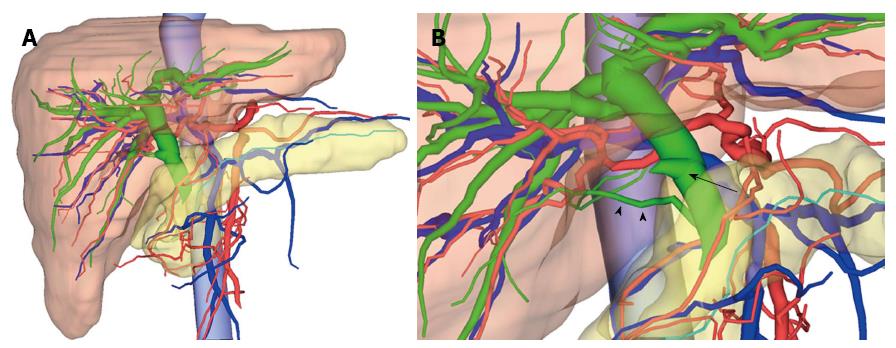
Figure 4 3-dimensional images by integrating multidetector computed tomography and magnetic resonance cholangiopancreatography images.
A: 3-dimensional (3D) image view from the front side of the patient. The red colour represents the arteries, the blue represents the veins and the portal vein, the green represents the biliary duct, and the turquoise represents the pancreatic duct. We were able to observe that the common and intrahepatic bile ducts were dilated; B: From the 3D image view from the patient's right side, the accessory bile duct from the caudate lobe connecting to the intrapancreatic bile duct (arrowheads) was easily recognisable. The cystic duct (arrow) has branched from the middle bile duct. We were able to determine that the injured site was the accessory bile duct from the caudate lobe.
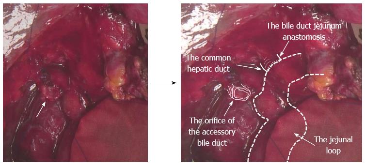
Figure 5 Intraoperative findings in the second operation.
The orifice of the injured caudate lobe bile duct of the proximal side on the surface of the liver was recognised (arrow). The precise anatomy is described using the annotation method.
- Citation: Miyamoto R, Oshiro Y, Hashimoto S, Kohno K, Fukunaga K, Oda T, Ohkohchi N. Three-dimensional imaging identified the accessory bile duct in a patient with cholangiocarcinoma. World J Gastroenterol 2014; 20(32): 11451-11455
- URL: https://www.wjgnet.com/1007-9327/full/v20/i32/11451.htm
- DOI: https://dx.doi.org/10.3748/wjg.v20.i32.11451


