Copyright
©2014 Baishideng Publishing Group Inc.
World J Gastroenterol. Aug 28, 2014; 20(32): 11443-11450
Published online Aug 28, 2014. doi: 10.3748/wjg.v20.i32.11443
Published online Aug 28, 2014. doi: 10.3748/wjg.v20.i32.11443
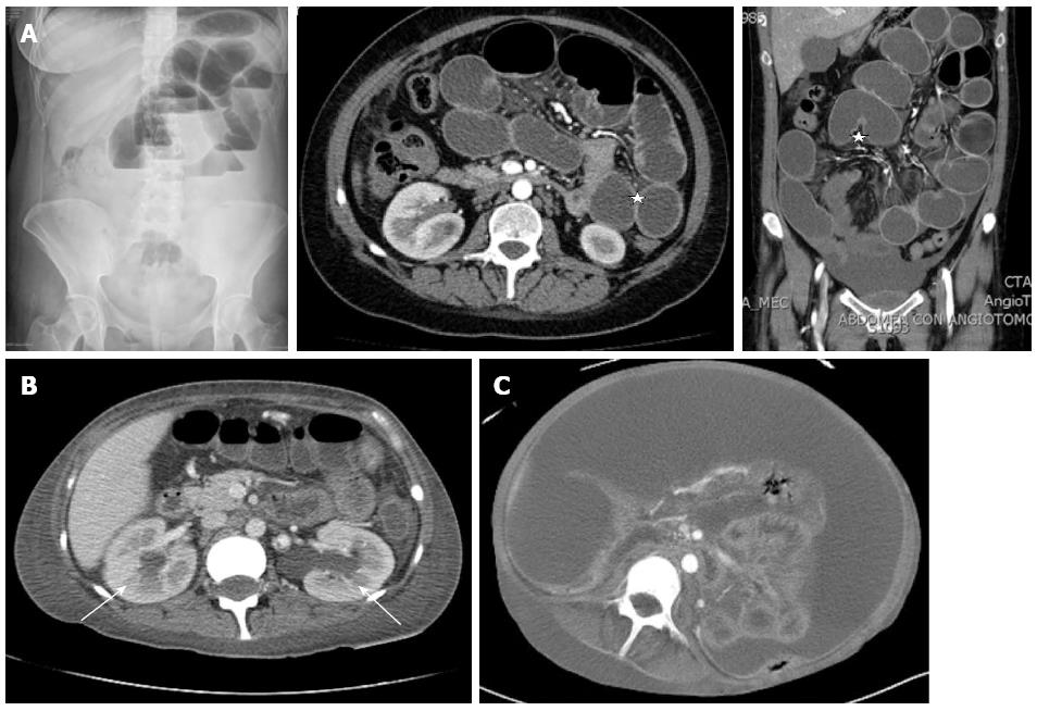
Figure 1 Imaging studies of intestinal pseudo-obstruction in lupus.
A: Represents the images where signs such as distention of the small bowel loops and air fluid levels are visible and edema of the wall labeled with stars known as target lesion; B: Shows the ureterohydronephrosis with labeled arrows (in this case bilateral), which is an accompanying frequent finding; C: Represents the image of intestinal visceromegaly.
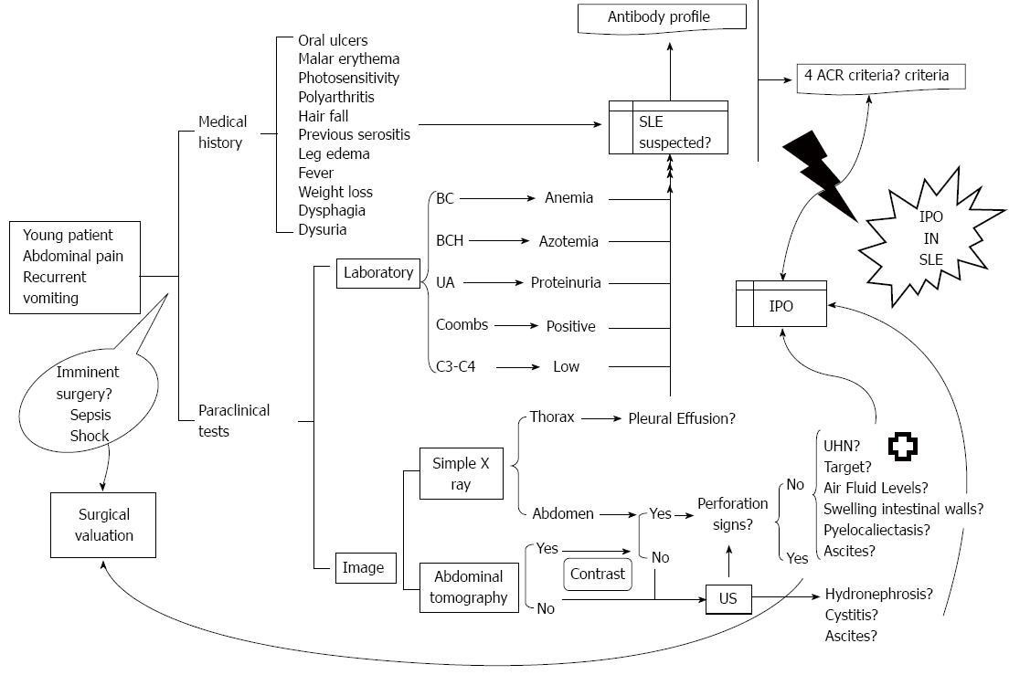
Figure 2 Suggested approach of intestinal pseudo-obstruction in lupus.
When a young patient presents with a clinical-radiological picture of intestinal pseudo-obstruction, the need for urgent surgery must be ruled out at first instance. Extensive interrogation and thorough examination must be carried out along with information that could suggest the picture. Among the initial examinations are X-ray, contrast abdominal tomography if possible or renal ultrasound if not possible, along with blood and urine tests. If information is compatible with the disease, specific serology should be carried out to determine if disease criteria have been met. If the diagnosis is corroborated, the patient should be evaluated by a specialist in order to initiate timely treatment. AA: Acute abdomen; BC: Blood cytometry; BCH: Blood chemistry; UA: Urinalysis; C3-C4: Complement; US: Ultrasound; IPO: Intestinal pseudo-obstruction; SLE: Systemic lupus eythematosus; ACR: American College of Rheumatology; UHN: Ureterohydronephrosis.
- Citation: López CAG, Laredo-Sánchez F, Malagón-Rangel J, Flores-Padilla MG, Nellen-Hummel H. Intestinal pseudo-obstruction in patients with systemic lupus erythematosus: A real diagnostic challenge. World J Gastroenterol 2014; 20(32): 11443-11450
- URL: https://www.wjgnet.com/1007-9327/full/v20/i32/11443.htm
- DOI: https://dx.doi.org/10.3748/wjg.v20.i32.11443


