Copyright
©2014 Baishideng Publishing Group Inc.
World J Gastroenterol. Aug 28, 2014; 20(32): 11376-11383
Published online Aug 28, 2014. doi: 10.3748/wjg.v20.i32.11376
Published online Aug 28, 2014. doi: 10.3748/wjg.v20.i32.11376
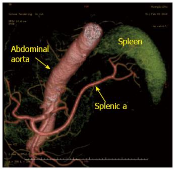
Figure 1 Preoperative computed tomography angiography showing the drainage of the splenic arteries.
Abdominal aorta (arrow); splenic a (arrow). a: Artery.
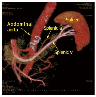
Figure 2 Preoperative computed tomography angiography showing the drainage of the splenic veins.
Abdominal aorta (arrow); splenic a (arrow); splenic v (arrow). a: Artery; v: Vein.
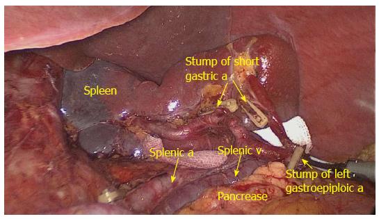
Figure 3 No.
10 lymph nodes lymphadenectomy at the front of the splenic vessels (anterior view). Dividing left gastroepiploic a (arrow); dividing short gastric a (arrow); splenic a (arrow); splenic v (arrow); a: Artery; v: Vein.
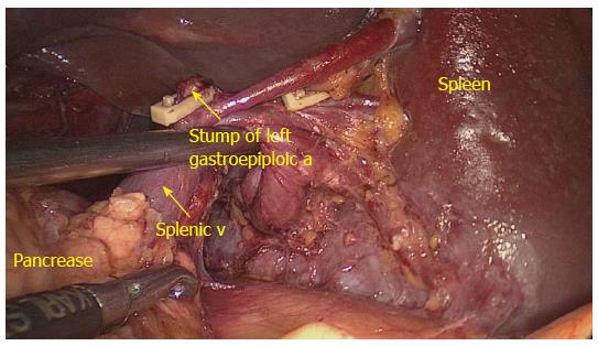
Figure 4 No.
10 lymph nodes lymphadenectomy behind the splenic vessels (posterior view). Dividing left gastroepiploic a (arrow); splenic vein (arrow); a: Artery; v: Vein.
- Citation: Li P, Huang CM, Zheng CH, Xie JW, Wang JB, Lin JX, Lu J, Wang Y, Chen QY. Laparoscopic spleen-preserving splenic hilar lymphadenectomy in 108 consecutive patients with upper gastric cancer. World J Gastroenterol 2014; 20(32): 11376-11383
- URL: https://www.wjgnet.com/1007-9327/full/v20/i32/11376.htm
- DOI: https://dx.doi.org/10.3748/wjg.v20.i32.11376


