Copyright
©2014 Baishideng Publishing Group Inc.
World J Gastroenterol. Jul 28, 2014; 20(28): 9476-9485
Published online Jul 28, 2014. doi: 10.3748/wjg.v20.i28.9476
Published online Jul 28, 2014. doi: 10.3748/wjg.v20.i28.9476
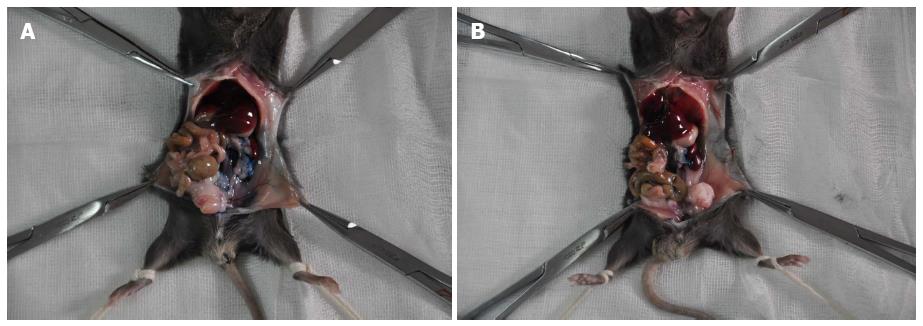
Figure 1 Pictorial depiction of the leakage of two different materials from the injection site.
Methylene blue containing phosphate buffered saline (A) and Matrigel (B) were injected into the parenchyma of the pancreatic tail of C57BL/6 mice.
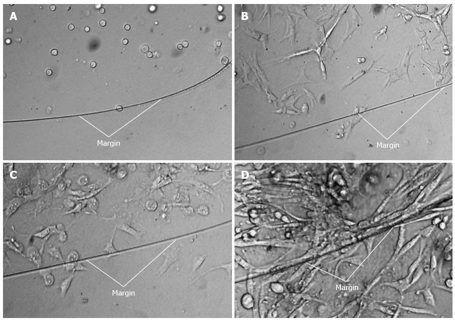
Figure 2 Adherence, proliferation, and expansion of Pan02 cells suspended in Matrigel and cultured ex vivo.
(A) 0 d, (B)1 d, (C) 2 d, and (D) 4 d. White lines indicated the margin of the Matrigel drip.
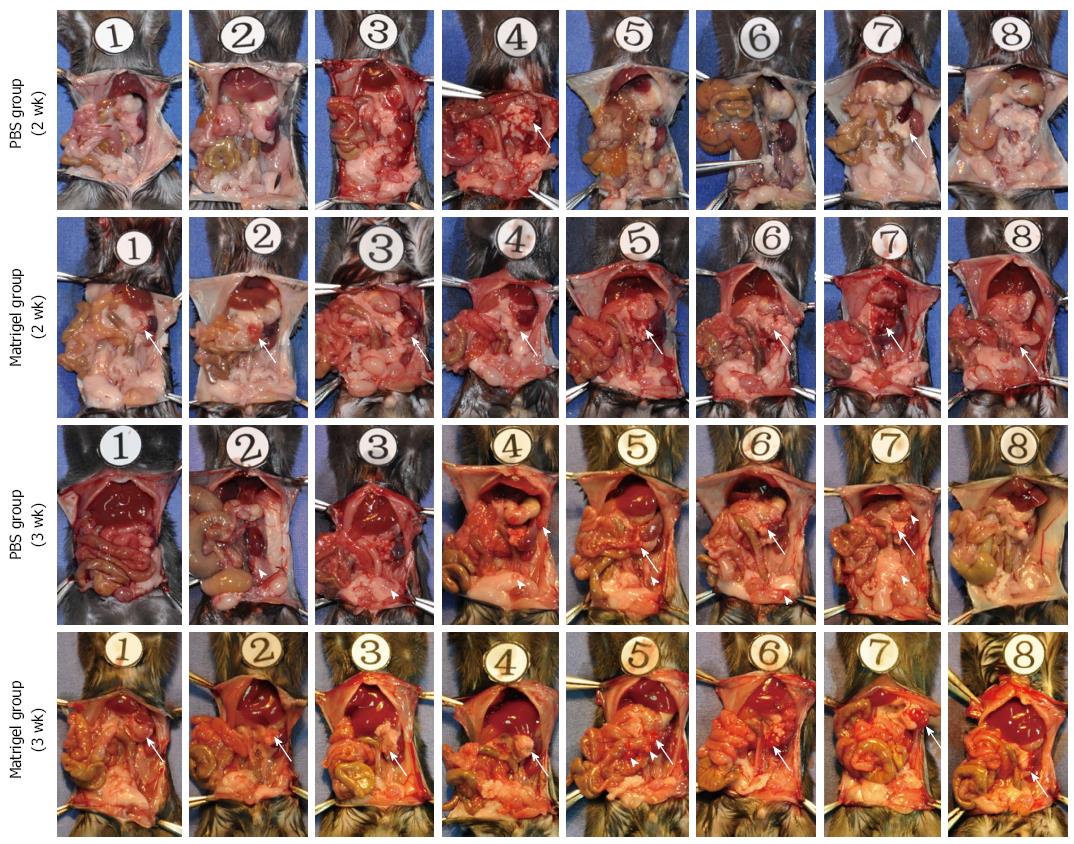
Figure 3 Tumor formation and intra-abdominal tumor implantation.
Pan02 cells were suspended in low-temperature phosphate buffered saline (PBS) or Matrigel, and injected into the pancreas tail of C57BL/6 mice. Half of the mice were sacrificed at 2 wk, with the remainder being sacrificed at 3 wk after injection. Tumor formation (white arrow) at the injection site and intra-abdominal tumor disseminated foci (white arrowhead) were thoroughly examined and marked.
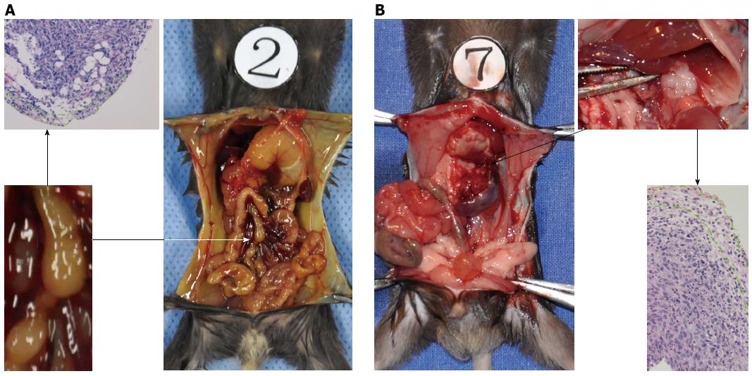
Figure 4 Different lymph node metastatic pathways arise from different sites of orthotopic pancreatic cancer.
Pancreatic cancer in the head (A) and pancreatic cancer in the tail (B). The region outlined by the green dot displayed residual lymphocytes in the metastatic lymph nodes.
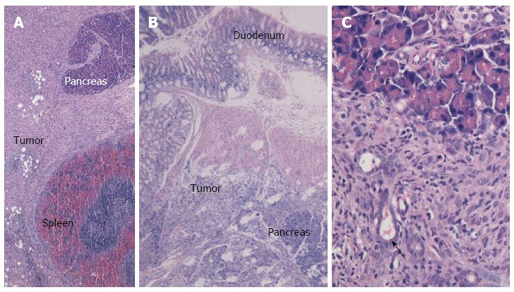
Figure 5 Cross-section and HE staining of orthotopic pancreatic cancer in the Matrigel group.
A: Full view of the pancreatic tail cancer (× 40); B: Full view of the pancreatic head cancer (× 40); C: Local view illustrating the duct-shape structure within the tumor tissue (× 200).
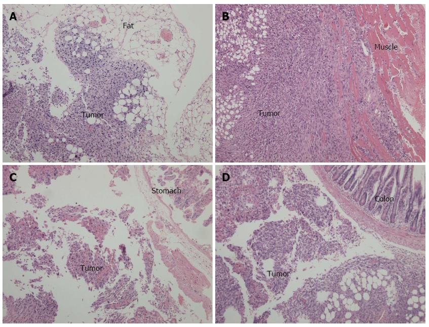
Figure 6 Cross-sections and hematoxylin-eosin staining of intra-abdominal disseminated foci observed under light microscopy (× 100).
A: Tumor implanted in the fat tissue; B: Tumor implanted in the abdominal wall; C: Tumor implanted in the stomach wall; D: Tumor implanted in the colon serosal.
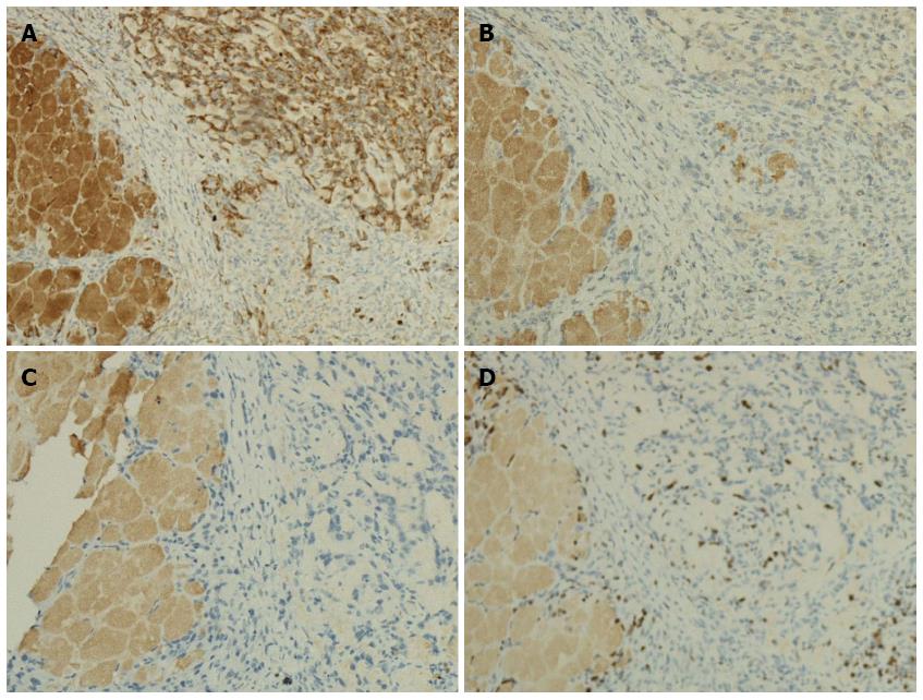
Figure 7 Immunohistochemical staining of Pan02 tumor sections, observed under light microscopy (× 100).
A: immunohistochemical staining of cytokeratin (yellow stained membrane indicates cytokeratin-positive cells); B: Immunohistochemical staining of vimentin; C: Immunohistochemical staining of chromogranin A; D: Immunohistochemical staining of Ki67 (yellow stained nuclei indicate Ki67-positive cells). The left portions of the sections are normal pancreatic tissue.
- Citation: Jiang YJ, Lee CL, Wang Q, Zhou ZW, Yang F, Jin C, Fu DL. Establishment of an orthotopic pancreatic cancer mouse model: Cells suspended and injected in Matrigel. World J Gastroenterol 2014; 20(28): 9476-9485
- URL: https://www.wjgnet.com/1007-9327/full/v20/i28/9476.htm
- DOI: https://dx.doi.org/10.3748/wjg.v20.i28.9476


