Copyright
©2014 Baishideng Publishing Group Inc.
World J Gastroenterol. Jun 28, 2014; 20(24): 7988-7992
Published online Jun 28, 2014. doi: 10.3748/wjg.v20.i24.7988
Published online Jun 28, 2014. doi: 10.3748/wjg.v20.i24.7988
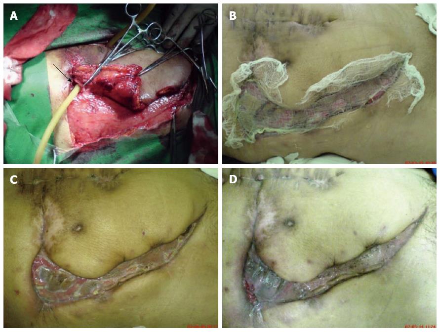
Figure 1 Debridement and drainage of fistula (arrow) (A); fistula healed and skin graft on wound (B); skin graft survived and grew (C); and the wound gradually healed (D).
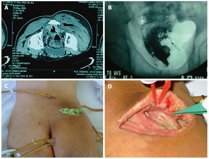
Figure 2 Computed tomography scan showing that the infection of the right hip had close relationship with rectum (arrow) (A); barium enema examination showed the fistula toward the right hip (arrow) (B); and placement of incisions and drainage tubes (C); and debridement and dressing change (D).
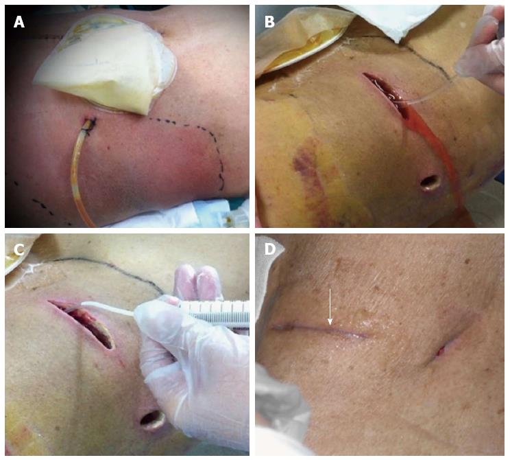
Figure 3 Inflamed area of the abdominal wall and parastomal fistula (supine position) (A); rinsing the wound with saline solution (right lateral position) (B); and infusing the wound with moist exposed burn ointment (syringe) (C); and the wound finally healed well (arrow) (D).
- Citation: Gu GL, Wang L, Wei XM, Li M, Zhang J. Necrotizing fasciitis secondary to enterocutaneous fistula: Three case reports. World J Gastroenterol 2014; 20(24): 7988-7992
- URL: https://www.wjgnet.com/1007-9327/full/v20/i24/7988.htm
- DOI: https://dx.doi.org/10.3748/wjg.v20.i24.7988


