Copyright
©2014 Baishideng Publishing Group Inc.
World J Gastroenterol. Jun 28, 2014; 20(24): 7777-7784
Published online Jun 28, 2014. doi: 10.3748/wjg.v20.i24.7777
Published online Jun 28, 2014. doi: 10.3748/wjg.v20.i24.7777
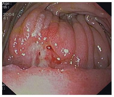
Figure 1 Patient with late hemorrhage after Roux-en-Y gastric bypass: endoscopic view shows a marginal ulceration with a visible vessel at the base of the lesion.
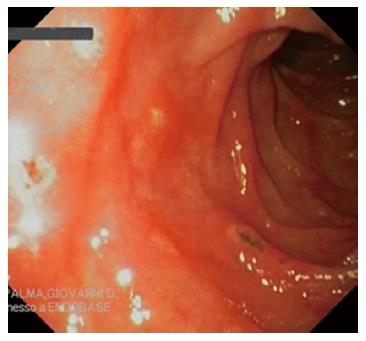
Figure 2 Multiple erosions in the anastomosed jejunum.
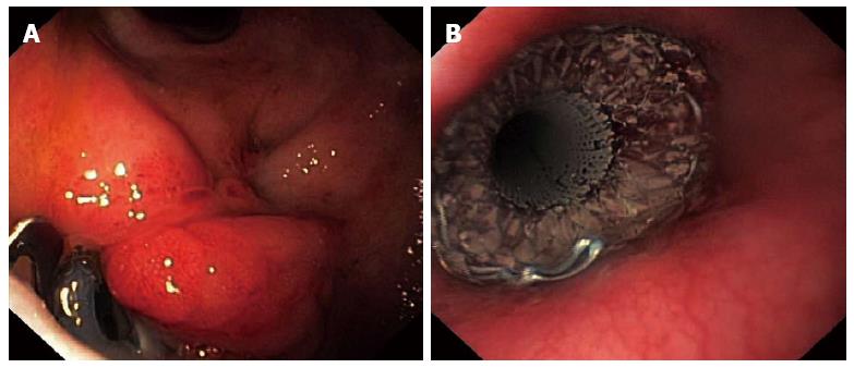
Figure 3 Patient with fistula after sleeve gastrectomy.
A: Endoscopic view of fistula at the upper portion of staple line in a patient after laparoscopic sleeve gastrectomy; a clip is visible on the boundary of the fistulous orifice as a result of a unsuccessful previous attempt at treatment; B: Fully covered removable stent (Taewoong Niti-S™ esophageal Mega stent) was implanted, promoting healing of the fistula.
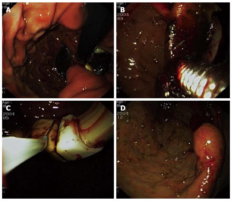
Figure 4 Different steps of endoscopic Lap-Band extraction.
A: Endoscopic view of an almost penetrated band, with only a small tissue bridge holding the device to the gastric wall; B: Endoscopic view of the AMI gastric band cutter (CJ Medical, Haddenham, United Kingdom) passed around the band after endoscopic resection of the tissue bridge; C: After the section, the band is grasped at the connection with the port-site, and extracted through the mouth; D: Gastric pouch outlet aspect after ring extraction (retrovision from the stomach).
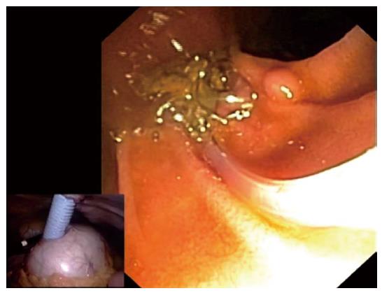
Figure 5 Laparoscopic assisted endoscopic retrograde cholangiopancreatography after Roux-en-Y gastric bypass.
A side-viewing endoscope is inserted through a 15-mm trocar that was previously placed into the excluded stomach. Subsequently, endoscopic retrograde cholangiopancreatography is performed in the usual fashion.
- Citation: De Palma GD, Forestieri P. Role of endoscopy in the bariatric surgery of patients. World J Gastroenterol 2014; 20(24): 7777-7784
- URL: https://www.wjgnet.com/1007-9327/full/v20/i24/7777.htm
- DOI: https://dx.doi.org/10.3748/wjg.v20.i24.7777


