Copyright
©2014 Baishideng Publishing Group Inc.
World J Gastroenterol. Jun 14, 2014; 20(22): 6897-6905
Published online Jun 14, 2014. doi: 10.3748/wjg.v20.i22.6897
Published online Jun 14, 2014. doi: 10.3748/wjg.v20.i22.6897
Figure 1 Changes in the body weight and daily food intake between hypothalamic nucleus-lesioned and sham-operated rats.
A: Body weight on days 3, 7, 14, and 21 post the operation; B: Daily food intake on days 3, 7, 14, and 21 post the operation. VMH: Ventromedial hypothalamic nucleus; VLH: Ventrolateral hypothalamic nucleus.
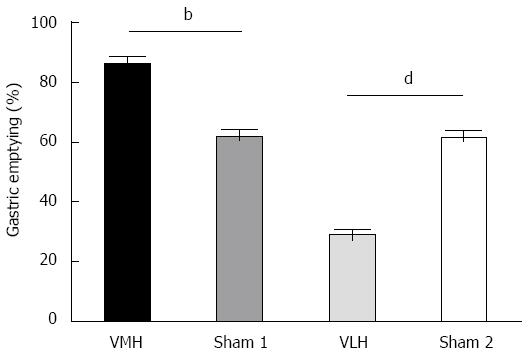
Figure 2 Effects of ventromedial hypothalamic nucleus and ventrolateral hypothalamic nucleus lesions on gastric emptying rate in rats.
The values represent mean ± SE (n = 6); bP < 0.01 and dP < 0.01 vs sham-operated rats. VMH: Ventromedial hypothalamic nucleus; VLH: Ventrolateral hypothalamic nucleus.
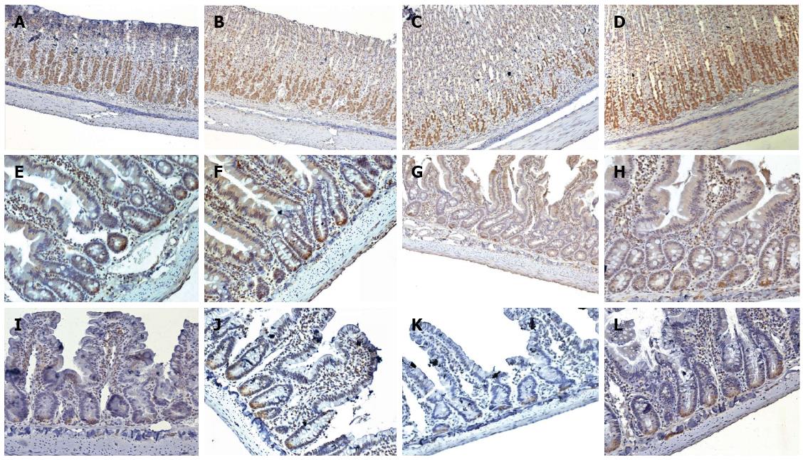
Figure 3 Immunohistochemical localisation of nesfatin-1 in the gastrointestinal tissues.
A-D: Nesfatin-1 IR cells (brown) in the stomach of VMH-lesioned, VMH-sham, VLH-lesioned, and VLH-sham rats, respectively; E-H: Nesfatin-1 IR cells (brown) in the duodenum of VMH-lesioned, VMH-sham, VLH-lesioned, and VLH-sham rats, respectively; I-L: Nesfatin-1 IR cells (brown) in the small intestine of VMH-lesioned, VMH-sham, VLH-lesioned, and VLH-sham rats, respectively. All of the magnifications are × 200 with the exception of the stomach, which is × 100. VMH: Ventromedial hypothalamic nucleus; VLH: Ventrolateral hypothalamic nucleus; IR: Immunoreactive.
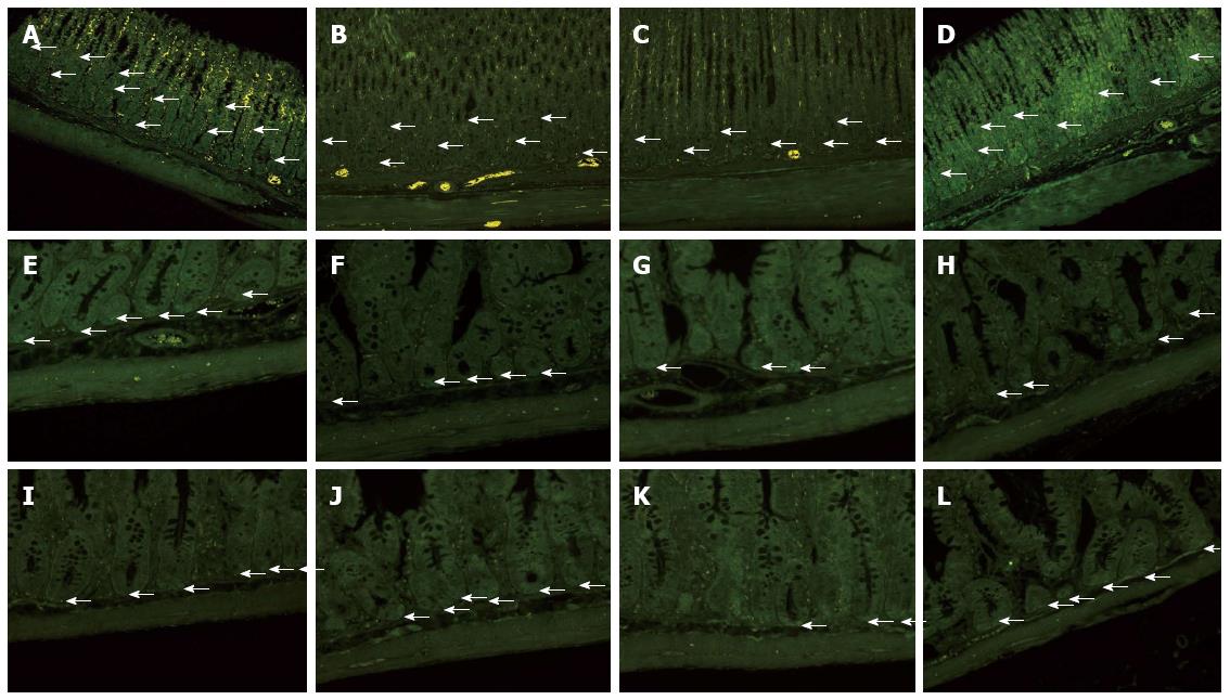
Figure 4 Immunofluorescence localisation of nesfatin-1 in the gastrointestinal tissue.
A-D: Nesfatin-1 IR cells (arrow; enhanced green) in the stomach of VMH-lesioned, VMH-sham, VLH-lesioned, and VLH-sham rats, respectively; E-H: Nesfatin-1 IR cells (arrow; enhanced green) in the duodenum of VMH-lesioned, VMH-sham, VLH-lesioned, and VLH-sham rats, respectively; I-L: Nesfatin-1 IR cells (arrow; enhanced green) in the small intestine of VMH-lesioned, VMH-sham, VLH-lesioned, and VLH-sham rats, respectively. All of the magnifications are × 200 with the exception of the stomach, which is × 100. VMH: Ventromedial hypothalamic nucleus; VLH: Ventrolateral hypothalamic nucleus; IR: Immunoreactive.
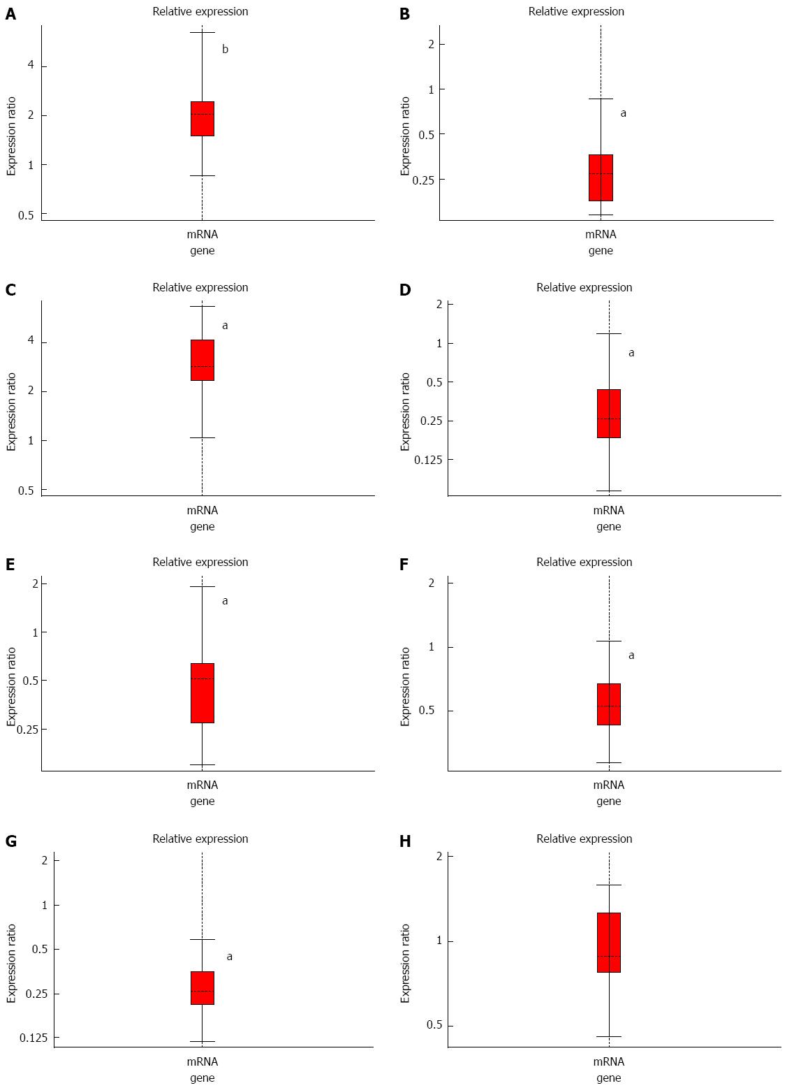
Figure 5 Relative expression levels of nucleobindin-2 mRNA in the stomach (A, B), duodenum (C, D), small intestine (E, F), and colon (G, H) between ventromedial hypothalamic nucleus- or ventrolateral hypothalamic nucleus-lesioned rats and the respective sham-operated rats.
Each tissue represents mean ± SE in the model rats and control groups (n = 6); bP < 0.01, aP < 0.05 vs sham-operated rats. VMH: Ventromedial hypothalamic nucleus; VLH: Ventrolateral hypothalamic nucleus.
- Citation: Tian ZB, Deng RJ, Sun GR, Wei LZ, Kong XJ, Ding XL, Jing X, Zhang CP, Ge YL. Expression of gastrointestinal nesfatin-1 and gastric emptying in ventromedial hypothalamic nucleus- and ventrolateral hypothalamic nucleus-lesioned rats. World J Gastroenterol 2014; 20(22): 6897-6905
- URL: https://www.wjgnet.com/1007-9327/full/v20/i22/6897.htm
- DOI: https://dx.doi.org/10.3748/wjg.v20.i22.6897


