Copyright
©2014 Baishideng Publishing Group Inc.
World J Gastroenterol. Jun 7, 2014; 20(21): 6541-6546
Published online Jun 7, 2014. doi: 10.3748/wjg.v20.i21.6541
Published online Jun 7, 2014. doi: 10.3748/wjg.v20.i21.6541
Figure 1 Esophagoduodenal anastomosis model: Esophagoduodenal anastomosis with total gastrectomy.
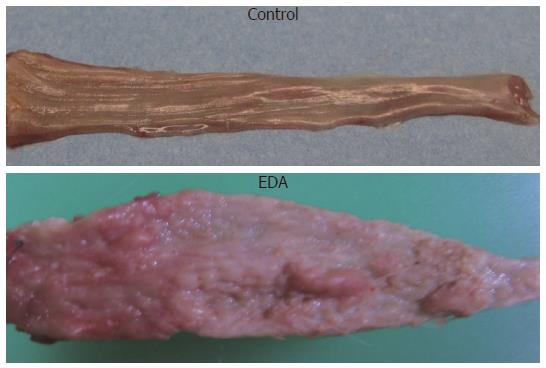
Figure 2 Macroscopic appearance of resected esophagi from esophagoduodenal anastomosis and control rats.
EDA: Esophagoduodenal anastomosis.
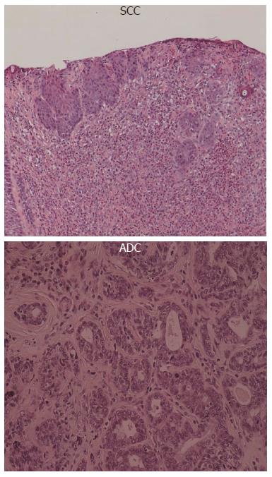
Figure 3 Microscopic findings in the distal portion of the esophagus from esophagoduodenal anastomosis rats.
ADC: Adenocarcinoma; SCC: Squamous cell carcinoma.
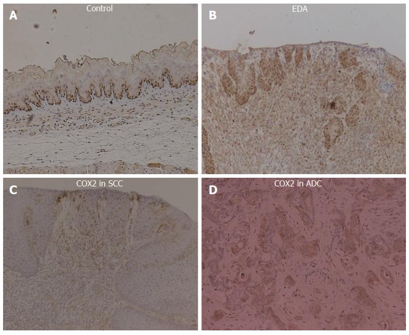
Figure 4 Immunohistochemical findings.
A: For proliferating cell nuclear antigen in control; B: Esophagoduodenal anastomosis (EDA) rats; C: For cyclooxygenase-2 (COX2) in squamous cell carcinoma (SCC); D: Adenocarcinoma (ADC) in EDA rats.
- Citation: Hashimoto N. Effects of bile acids on cyclooxygenase-2 expression in a rat model of duodenoesophageal anastomosis. World J Gastroenterol 2014; 20(21): 6541-6546
- URL: https://www.wjgnet.com/1007-9327/full/v20/i21/6541.htm
- DOI: https://dx.doi.org/10.3748/wjg.v20.i21.6541


