Copyright
©2014 Baishideng Publishing Group Co.
World J Gastroenterol. Jan 14, 2014; 20(2): 607-610
Published online Jan 14, 2014. doi: 10.3748/wjg.v20.i2.607
Published online Jan 14, 2014. doi: 10.3748/wjg.v20.i2.607
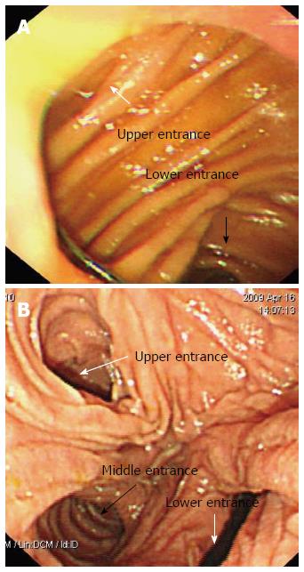
Figure 1 The gastrojejunal anastomosis is detected at the distal end of the stomach, and 2 stomal openings corresponding to an end-to-side anastomosis can be identified endoscopically.
If the efferent loop was constructed at the greater curvature of the stomach in the previous surgery, the “lower entrance” is the entrance to the right efferent loop (A). Three stomal openings can be identified endoscopically at the site of the Braun anastomosis, and the “middle entrance” leads to the appropriate loop to reach the papilla of Vater. The “middle entrance” is unique irrespective of the endoscopic approach used (B).
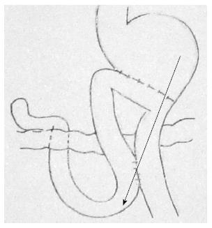
Figure 2 The duodenoscope should be extended along the greater curvature of the stomach and then advanced through the “lower entrance” at the site of the gastrojejunal anastomosis, along the efferent loop, and through the “middle entrance” at the site of the Braun anastomosis to reach the papilla of Vater.
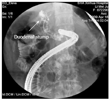
Figure 3 Retrieval-balloon-assisted enterography.
A catheter is advanced into the middle limb and contrast injected into the loop to confirm that the limb is the duodenal stump.
- Citation: Wu WG, Gu J, Zhang WJ, Zhao MN, Zhuang M, Tao YJ, Liu YB, Wang XF. ERCP for patients who have undergone Billroth II gastroenterostomy and Braun anastomosis. World J Gastroenterol 2014; 20(2): 607-610
- URL: https://www.wjgnet.com/1007-9327/full/v20/i2/607.htm
- DOI: https://dx.doi.org/10.3748/wjg.v20.i2.607


