Copyright
©2014 Baishideng Publishing Group Co.
World J Gastroenterol. Jan 14, 2014; 20(2): 555-560
Published online Jan 14, 2014. doi: 10.3748/wjg.v20.i2.555
Published online Jan 14, 2014. doi: 10.3748/wjg.v20.i2.555
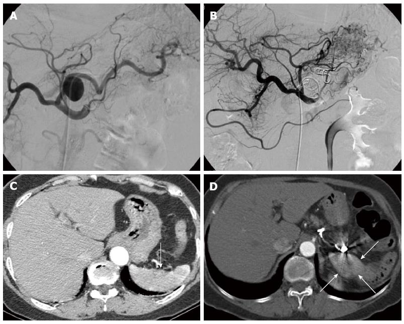
Figure 1 Group A.
A splenic artery aneurysm (SAA) in a 56-year-old man with normal spleen. A: Celiac arteriogram prior to coil embolization demonstrated an SAA located at the proximal splenic artery; B: Celiac arteriogram after coil embolization showed total embolization of the main splenic artery with disappearance of the SAA; C: Before embolization, arterial phase of computed tomography angiography (CTA) showed that the spleen was normal (arrow); D: Arterial phase of CTA showed that the spleen had decreased size (arrows) 2 years after embolization.
- Citation: Li ES, Mu JX, Ji SM, Li XM, Xu LB, Chai TC, Liu JX. Total splenic artery embolization for splenic artery aneurysms in patients with normal spleen. World J Gastroenterol 2014; 20(2): 555-560
- URL: https://www.wjgnet.com/1007-9327/full/v20/i2/555.htm
- DOI: https://dx.doi.org/10.3748/wjg.v20.i2.555


