Copyright
©2014 Baishideng Publishing Group Co.
World J Gastroenterol. Jan 14, 2014; 20(2): 436-444
Published online Jan 14, 2014. doi: 10.3748/wjg.v20.i2.436
Published online Jan 14, 2014. doi: 10.3748/wjg.v20.i2.436
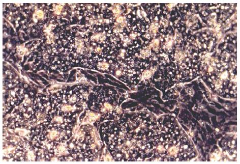
Figure 1 Primary duck embryonic hepatocytes grown in CeLLBINDTM plates at day 3 of cultivation.
Monolayers of hepatocytes, which show typical polygonal morphology, are interrupted by areas of non-parenchymal cells (light microscopy, phase contrast, x 200).
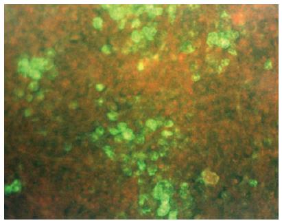
Figure 2 Detection of duck hepatitis B virus-specific surface antigen six days after inoculation of primary duck embryonic hepatocytes by indirect immunofluorescence.
Polyvalent rabbit anti-DHBs (kindly provided by Dr. D. Glebe, Institute of Medical Virology, National Reference Centre for Hepatitis B and D, Justus Liebig University, Giessen, Germany) and goat anti-rabbit IgG Alexa Fluor® 488 (Life Technologies, Darmstadt, Germany) were used as antibodies (fluorescence microscopy, x 125).
- Citation: Sauerbrei A. Is hepatitis B-virucidal validation of biocides possible with the use of surrogates? World J Gastroenterol 2014; 20(2): 436-444
- URL: https://www.wjgnet.com/1007-9327/full/v20/i2/436.htm
- DOI: https://dx.doi.org/10.3748/wjg.v20.i2.436


