Copyright
©2014 Baishideng Publishing Group Co.
World J Gastroenterol. May 14, 2014; 20(18): 5461-5473
Published online May 14, 2014. doi: 10.3748/wjg.v20.i18.5461
Published online May 14, 2014. doi: 10.3748/wjg.v20.i18.5461
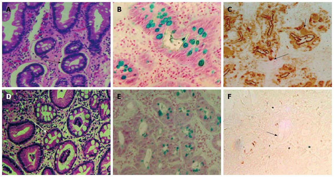
Figure 1 Serial sections of formalin fixed paraffin embedded biopsy tissue from two patients with gastric intestinal metaplasia without carcinoma.
Haematoxylin-eosin staining (A, D), alcian blue/high iron diamine staining (B, E), and immunoperoxidase assay with the monoclonal antibody mAb Das-1 (C, F); (A-C) is from the same patient and (D-F) from the second patient. mAb Das-1 stained both goblet cells (shorter arrow) and metaplastic non-goblet cells (longer arrow) in the glands (C), suggesting colonic phenotype (incomplete type); While GIM is clearly evident with the presence of goblet cells (D, E) in the second patient, but mAb Das-1 did not stain the glands including goblet cells (F). The arrow shows the unstained goblet cells suggesting complete phenotype or small intestinal phenotype (original magnification × 160 for each part)[106].
-
Citation: Watari J, Chen N, Amenta PS, Fukui H, Oshima T, Tomita T, Miwa H, Lim KJ, Das KM.
Helicobacter pylori associated chronic gastritis, clinical syndromes, precancerous lesions, and pathogenesis of gastric cancer development. World J Gastroenterol 2014; 20(18): 5461-5473 - URL: https://www.wjgnet.com/1007-9327/full/v20/i18/5461.htm
- DOI: https://dx.doi.org/10.3748/wjg.v20.i18.5461


