Copyright
©2014 Baishideng Publishing Group Co.
World J Gastroenterol. Apr 28, 2014; 20(16): 4817-4821
Published online Apr 28, 2014. doi: 10.3748/wjg.v20.i16.4817
Published online Apr 28, 2014. doi: 10.3748/wjg.v20.i16.4817
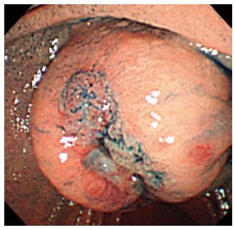
Figure 1 A submucosal tumor with central ulceration at the ampulla of Vater.
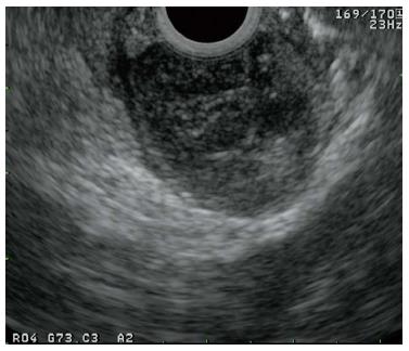
Figure 2 Endoscopic ultrasonography revealed a round, low-echoic mass.
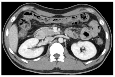
Figure 3 Enhanced computed tomography scan revealed a smooth-outlined hypervascular solid mass.
Figure 4 Mass showed low signal intensity on T1-weighted images (A) and high signal intensity on T2-weighted images (B).
Figure 5 Microscopic examination revealed a spindle-cell neoplasm (A, hematoxylin and eosin, × 200), and the tumor cells were positive for c-kit (B, × 200) and CD34 (C, × 200).
- Citation: Kobayashi M, Hirata N, Nakaji S, Shiratori T, Fujii H, Ishii E. Gastrointestinal stromal tumor of the ampulla of Vater: A case report. World J Gastroenterol 2014; 20(16): 4817-4821
- URL: https://www.wjgnet.com/1007-9327/full/v20/i16/4817.htm
- DOI: https://dx.doi.org/10.3748/wjg.v20.i16.4817


