Copyright
©2014 Baishideng Publishing Group Co.
World J Gastroenterol. Apr 28, 2014; 20(16): 4797-4805
Published online Apr 28, 2014. doi: 10.3748/wjg.v20.i16.4797
Published online Apr 28, 2014. doi: 10.3748/wjg.v20.i16.4797
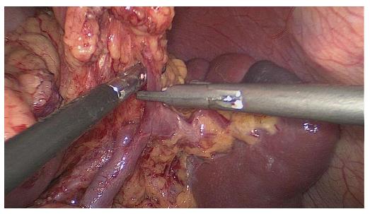
Figure 1 Left gastroepiploic vessels are vascularised at the origin.
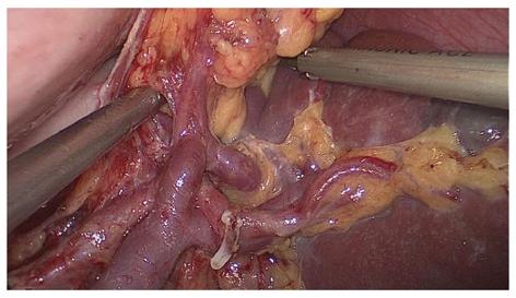
Figure 2 Short gastric vessels are freed, clamped, and cut at the origin.
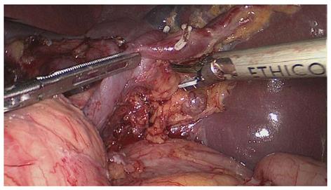
Figure 3 Lymphatic fatty tissues behind the splenic hilum are dissected.
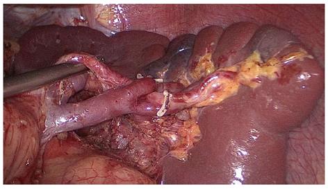
Figure 4 Fatty tissues, including the splenic lymph nodes, are en-bloc removed from the splenic hilum.
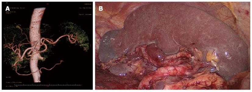
Figure 5 A single-lobe splenic vessel.
A: Preoperative assessment of the splenic vascular anatomy using computed tomography with 3D imaging images; B: Operative view after the completion of splenic lymph node dissection.
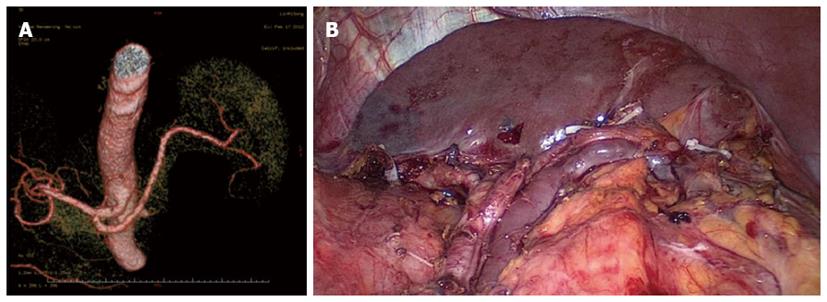
Figure 6 Two-lobe splenic vessels.
A: Preoperative assessment of the splenic vascular anatomy using computed tomography with 3D imaging images; B: Operative view after the completion of splenic lymph node dissection.
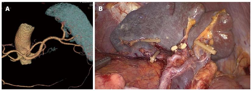
Figure 7 Three-lobe splenic vessels.
A: Preoperative assessment of the splenic vascular anatomy using computed tomography with 3D imaging images; B: Operative view after the completion of splenic lymph node dissection.
- Citation: Wang JB, Huang CM, Zheng CH, Li P, Xie JW, Lin JX, Lu J. Role of 3DCT in laparoscopic total gastrectomy with spleen-preserving splenic lymph node dissection. World J Gastroenterol 2014; 20(16): 4797-4805
- URL: https://www.wjgnet.com/1007-9327/full/v20/i16/4797.htm
- DOI: https://dx.doi.org/10.3748/wjg.v20.i16.4797


