Copyright
©2014 Baishideng Publishing Group Co.
World J Gastroenterol. Apr 28, 2014; 20(16): 4712-4717
Published online Apr 28, 2014. doi: 10.3748/wjg.v20.i16.4712
Published online Apr 28, 2014. doi: 10.3748/wjg.v20.i16.4712
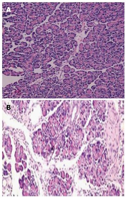
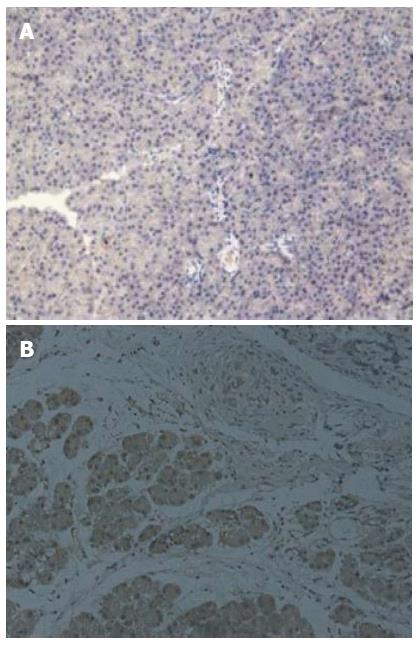
Figure 1 Histologic examination of pancreas (× 200).
A: Normal rats; B: Chronic pancreatitis rats. Dense inflammatory infiltrates, large regions of glandular atrophy, pseudo tubular complexes, edema, and fibrosis replaced the normal pancreas (B).
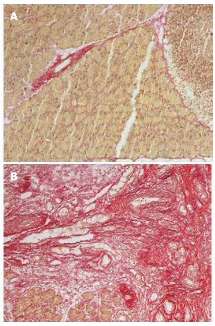
Figure 2 Sirius red staining of pancreas (× 200).
A: Normal rats; B: Chronic pancreatitis rats. Significantly higher expression of lobular and sublobular collagen deposition is seen in Figure B.
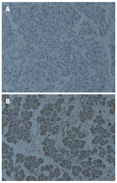
Figure 3 Immunohistochemical staining of Ptch1 in pancreas (× 200).
A: Normal rats; B: Chronic pancreatitis rats. Significantly higher expression of Ptch1 is seen in Figure B, and Ptch1 expression was mainly restricted to ductal, acinar, or islet compartments of the pancreas.
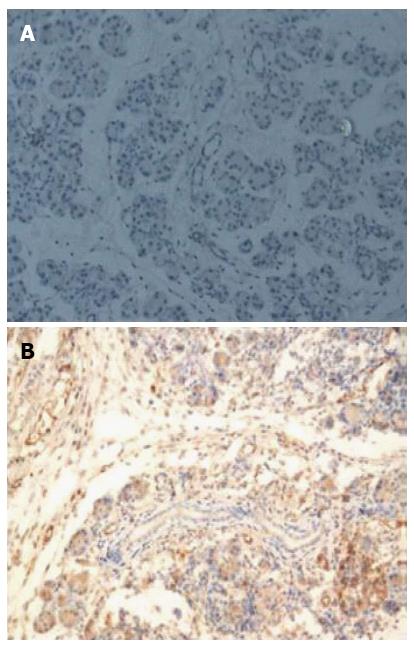
Figure 4 Immunohistochemical staining of Smo in pancreas (× 200).
A: Normal rats; B: Chronic pancreatitis rats. Significantly higher expression of Smo is seen in Figure 4B, and the location of Smo expression was mainly restricted to ductal, acinar, or islet compartments of the pancreas.
Figure 5 Immunohistochemical staining of Gli1 in pancreas (× 200).
A: Normal rats; B: Chronic pancreatitis rats. Significantly higher expression of Gli1 is seen in Figure 5B, and Gli1 expression was mainly restricted to ductal, acinar, or islet compartments of the pancreas.
- Citation: Wang LW, Lin H, Lu Y, Xia W, Gao J, Li ZS. Sonic hedgehog expression in a rat model of chronic pancreatitis. World J Gastroenterol 2014; 20(16): 4712-4717
- URL: https://www.wjgnet.com/1007-9327/full/v20/i16/4712.htm
- DOI: https://dx.doi.org/10.3748/wjg.v20.i16.4712


