Copyright
©2014 Baishideng Publishing Group Co.
World J Gastroenterol. Apr 28, 2014; 20(16): 4526-4535
Published online Apr 28, 2014. doi: 10.3748/wjg.v20.i16.4526
Published online Apr 28, 2014. doi: 10.3748/wjg.v20.i16.4526
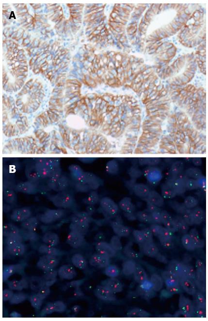
Figure 1 Human epidermal growth factor receptor 2 positive gastric adenocarcinoma.
A: Immunohistochemistry (HercepTest™, Dako); B: Fluorescence in situ hybridization (FISH) (Human epidermal growth factor receptor 2 FISH pharmDx™ Kit, Dako).
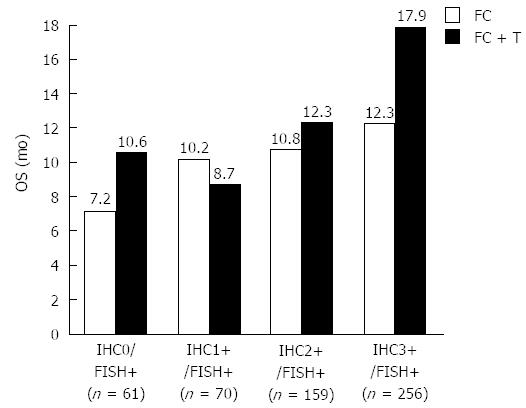
Figure 2 Median overall survival in months for the four individual human epidermal growth factor receptor 2 immunohistochemistry scores for the two treatment groups.
OS: Overall survival; FC: Fluorouracil/capecitabine plus cisplatin; FC + T: Fluorouracil/capecitabine plus cisplatin plus trastuzumab[10]; FISH: Fluorescence in situ hybridization; IHC: Immunohistochemistry.
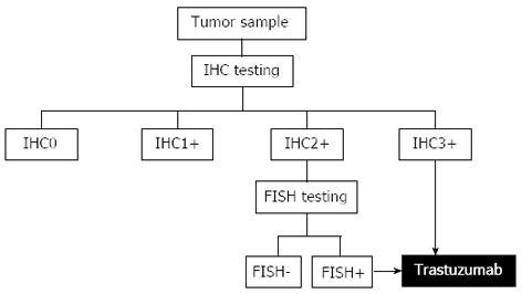
Figure 3 Human epidermal growth factor receptor 2 testing algorithm developed based on the results of the ToGA trial.
Immunohistochemistry (IHC) is the primary test with reflex testing with Fluorescence in situ hybridization (FISH) in case of an equivocal IHC result (IHC2+)[27].
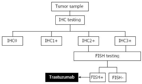
Figure 4 Human epidermal growth factor receptor 2 testing algorithm with fluorescence in situ hybridization reflex testing for both immunohistochemistry 2+ and immunohistochemistry 3+.
This testing algorithm was recommended by the United States Food and Drug Administration in relation to approval of trastuzumab for advanced gastric cancer. FISH: Fluorescence in situ hybridization; IHC: Immunohistochemistry.
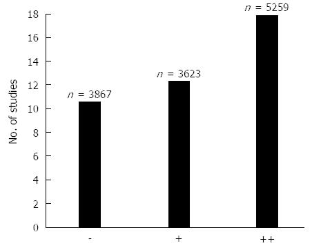
Figure 5 The number of studies and patients (n) in each of the three scoring categories.
Symbols: Two pluses (++) indicate the strongest association with the Human epidermal growth factor receptor 2 (HER2)+ status, one plus (+) indicates a somewhat weaker association with the HER2+ status, and minus (–) indicates that no associations with the HER2+ was found[6].
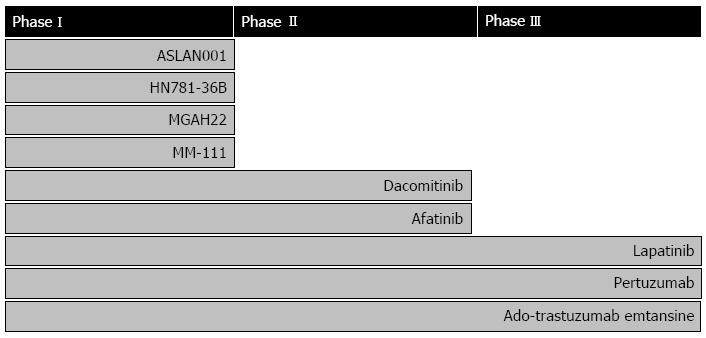
Figure 6 Drugs targeting human epidermal growth factor receptor 2 in clinical development for treatment of gastric, esophageal, or gastroesophageal junction cancer.
The individual compounds are listed according to the stage of development[35].
- Citation: Jørgensen JT. Role of human epidermal growth factor receptor 2 in gastric cancer: Biological and pharmacological aspects. World J Gastroenterol 2014; 20(16): 4526-4535
- URL: https://www.wjgnet.com/1007-9327/full/v20/i16/4526.htm
- DOI: https://dx.doi.org/10.3748/wjg.v20.i16.4526


