Copyright
©2014 Baishideng Publishing Group Co.
World J Gastroenterol. Mar 28, 2014; 20(12): 3245-3254
Published online Mar 28, 2014. doi: 10.3748/wjg.v20.i12.3245
Published online Mar 28, 2014. doi: 10.3748/wjg.v20.i12.3245
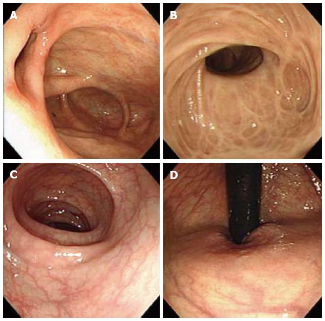
Figure 1 Colonoscopic findings at clinical onset.
A: Cecum; B: Ascending colon; C: Sigmoid colon; D: Rectum. A 43-year-old female patient diagnosed with asymptomatic, concurrent primary sclerosing cholangitis-inflammatory bowel disease. The first colonoscopy showed multiple white scars in the ascending colon and right-sided transverse colon and no abnormal findings in the left-sided transverse colon, descending colon, sigmoid colon, or rectum.
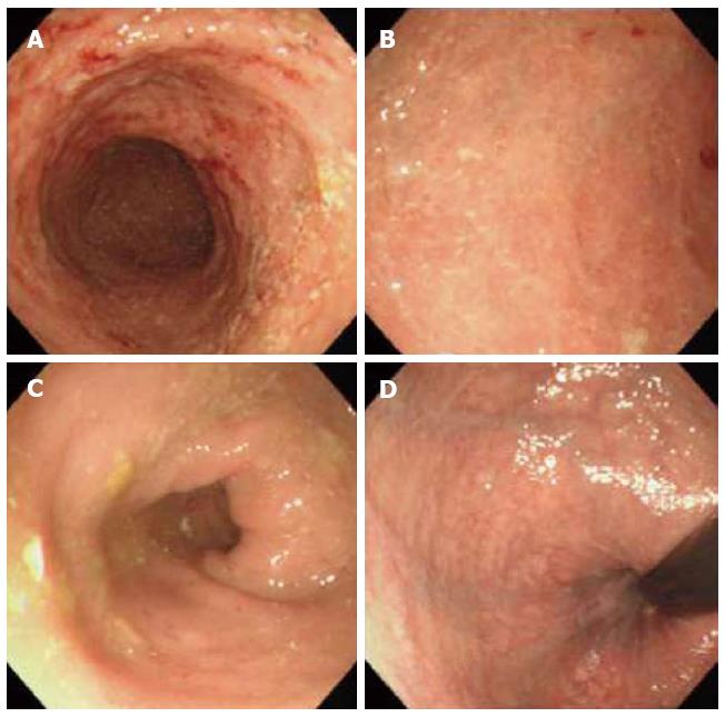
Figure 2 Colonoscopic findings seven months later.
A: Cecum; B: Ascending colon; C: Sigmoid colon; D: Rectum. A repeat colonoscopy seven months later showing inflamed mucosa with multiple erosions and redness from the ascending colon to the right-sided transverse colon. Mucosal vessels are clearly visible in the descending colon, sigmoid colon, and rectum.
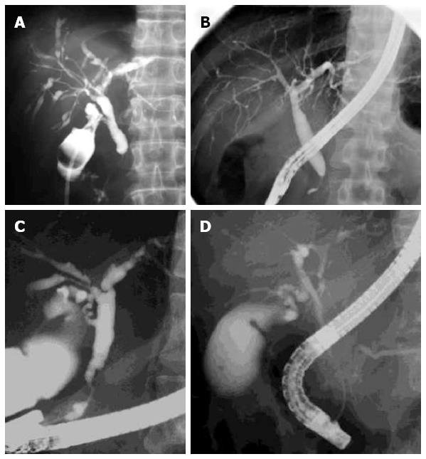
Figure 3 Cholangiographic examples of immunoglobulin G4-related sclerosing cholangitis and primary sclerosing cholangitis.
Cholangiograms of immunoglobulin G4-related sclerosing cholangitis showing multiple stenoses in the intrahepatic ducts and stenosis in the intrapancreatic portion (A, B). Cholangiograms of primary sclerosing cholangitis showing a beaded appearance (C) and pruning of the intrahepatic ducts (C, D).
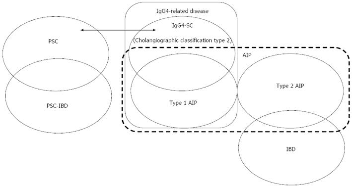
Figure 4 Correlation between inflammatory bowel disease and screrosing cholangitis.
PSC is frequently associated with characteristic PSC-IBD, whereas IgG4-SC is not associated with IBD. IgG4-SC is frequently associated with type 1 AIP, whereas type 2 AIP is frequently associated with IBD. PSC: Primary sclerosing cholangitis; PSC-IBD: IBD-associated with PSC; IBD: Inflammatory bowel disease; IgG4-SC: Immunoglobulin G4-related sclerosing cholangitis; AIP: Autoimmune pancreatitis.
- Citation: Nakazawa T, Naitoh I, Hayashi K, Sano H, Miyabe K, Shimizu S, Joh T. Inflammatory bowel disease of primary sclerosing cholangitis: A distinct entity? World J Gastroenterol 2014; 20(12): 3245-3254
- URL: https://www.wjgnet.com/1007-9327/full/v20/i12/3245.htm
- DOI: https://dx.doi.org/10.3748/wjg.v20.i12.3245


