Copyright
©2014 Baishideng Publishing Group Co.
World J Gastroenterol. Mar 14, 2014; 20(10): 2731-2734
Published online Mar 14, 2014. doi: 10.3748/wjg.v20.i10.2731
Published online Mar 14, 2014. doi: 10.3748/wjg.v20.i10.2731
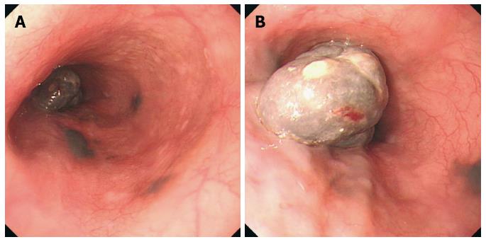
Figure 1 Endoscopic examination findings.
A: A large (20 mm × 40 mm) tumor in the esophagus 33 cm from the incisors and 3 pigmented spots (satellites) adjacent to the large tumor 30-33 cm from the incisors; B: A partial stalk and the black uneven surface of the large tumor and the dark smooth surface of the 3 flat lesions.
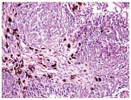
Figure 2 Pathological findings (hematoxylin/eosin staining) from the endoscopy.
The tumor contained variously pigmented bizarre cells.
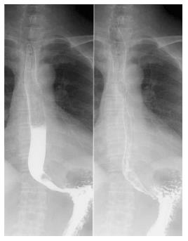
Figure 3 Upper gastrointestinal series showing a circular filling defect in the lower esophagus.
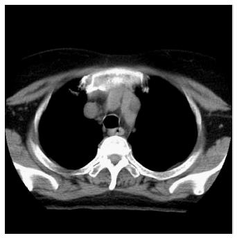
Figure 4 Plain computed tomography scan of the cervical region, chest and upper abdomen demonstrating the thickness of the middle esophagus wall.
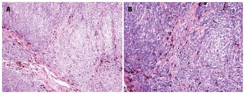
Figure 5 Pathological findings (hematoxylin/eosin staining) from the postoperative specimens.
A: Large tumor (× 100); B: Large tumor (× 200).
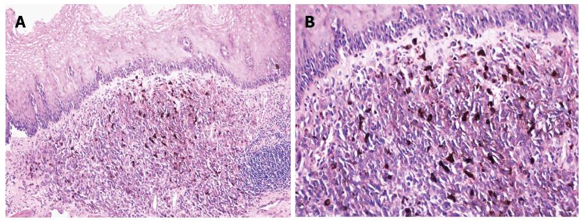
Figure 6 Pathological findings (hematoxylin/eosin staining) from the postoperative specimens.
A: One of the satellites (× 100); B: One of the satellites (× 200).
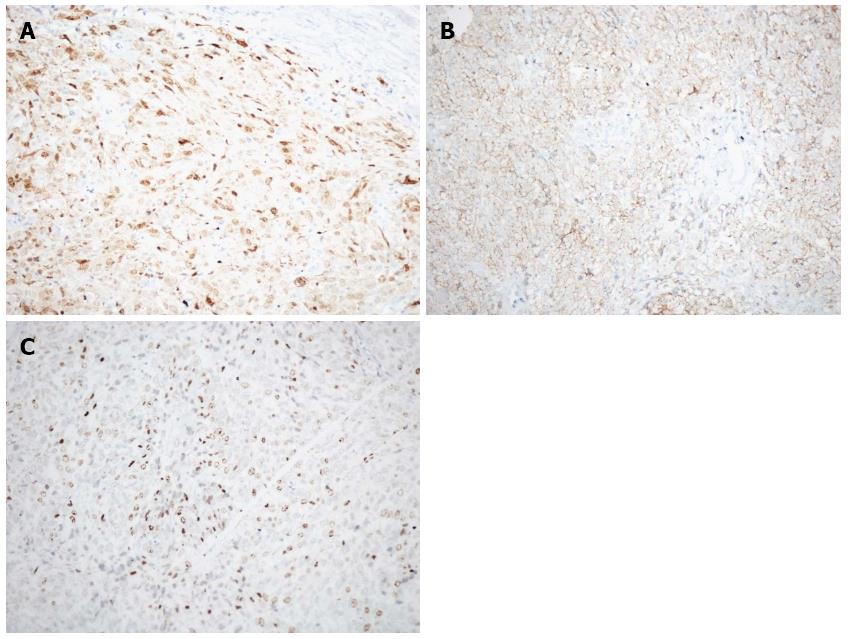
Figure 7 Immunohistochemical staining positive for S100 (A), HMB45 (B), and Ki67 (C).
- Citation: Li YH, Li X, Zou XP. Primary malignant melanoma of the esophagus: A case report. World J Gastroenterol 2014; 20(10): 2731-2734
- URL: https://www.wjgnet.com/1007-9327/full/v20/i10/2731.htm
- DOI: https://dx.doi.org/10.3748/wjg.v20.i10.2731


