Copyright
©The Author(s) 1996.
World J Gastroenterol. Jun 25, 1996; 2(2): 123-124
Published online Jun 25, 1996. doi: 10.3748/wjg.v2.i2.123
Published online Jun 25, 1996. doi: 10.3748/wjg.v2.i2.123
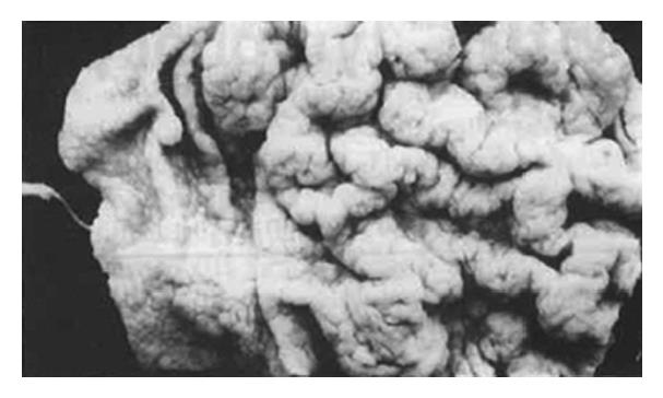
Figure 1 Mucosal folds of the body of the stomach with appearance reminiscent of cerebral convolution.
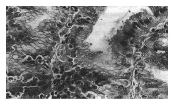
Figure 2 Numorous intraepithelial lymphocytic infiltration of surface and epithelial pits.
HE staining, magnification × 132.
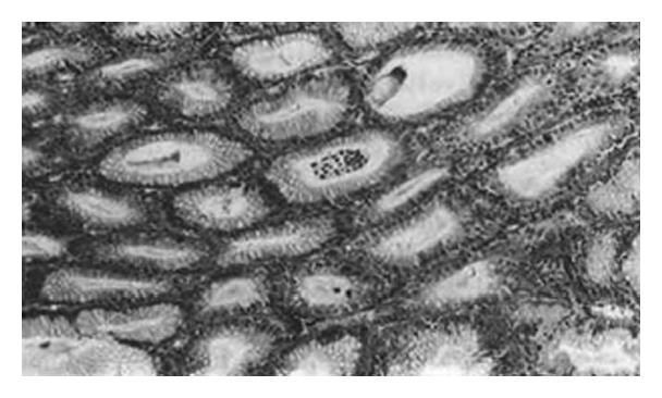
Figure 3 Crypts lined with mucus-secreting cells and containing lymphocytes and neutrophils, with formation of crypt abscesses.
HE staining, magnification × 66.
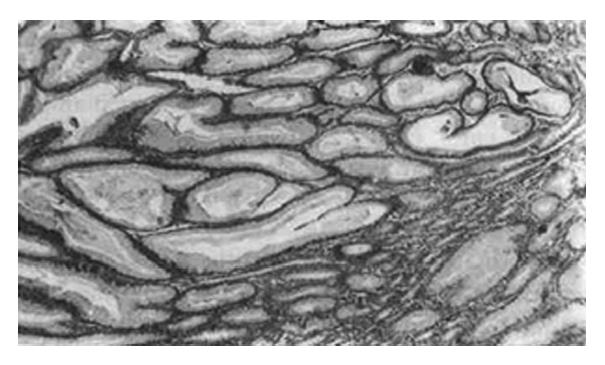
Figure 4 Gland normally containing parietal and chief cells that were replaced by the active mucus-secreting cells.
HE staining, magnification × 66.
- Citation: Zhang XS, Zhang Y, Wu SH, Han YZ, Li B, Ma Y. Menetrier’s disease with lymphocytic gastritis: Report of two cases. World J Gastroenterol 1996; 2(2): 123-124
- URL: https://www.wjgnet.com/1007-9327/full/v2/i2/123.htm
- DOI: https://dx.doi.org/10.3748/wjg.v2.i2.123


