Copyright
©2013 Baishideng Publishing Group Co.
World J Gastroenterol. Feb 28, 2013; 19(8): 1322-1326
Published online Feb 28, 2013. doi: 10.3748/wjg.v19.i8.1322
Published online Feb 28, 2013. doi: 10.3748/wjg.v19.i8.1322
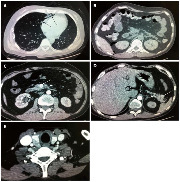
Figure 1 Computed tomography scan of the case.
A: Chest Computed tomography (CT) shows a mediastinal emphysema (arrows); B: Epigastric CT shows accumulation of gas and fluid (arrows) in the anterior pararenal space just adjacent to the thickened wall of the horizontal part of the duodenum as well as accumulation of gas in the right perirenal space; C, D: A small intestine CT shows bowel wall thickening and strong enhancement of the horizontal part of the duodenum and a nodular mass (arrows) with a rich blood supply in the uncinate process (C) and tail (D) of the pancreas; E: Thyroid gland CT shows mild nodular goiter with nodules (arrow) posterior and lateral to the thyroid gland.
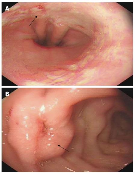
Figure 2 Gastroscopy reveals reflux esophagitis LA grade C and multiple deep ulcers in the descending part of duodenum, arrows indicate the erosion and ulcer respectively.
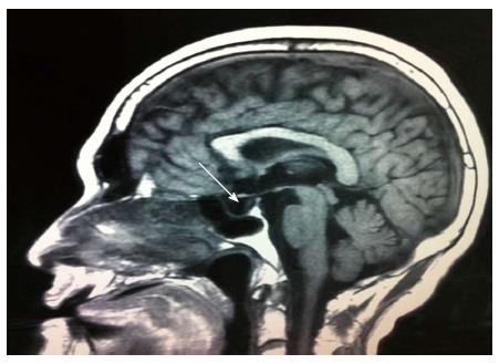
Figure 3 Pituitary Magnetic resonance imaging shows no space-occupying lesion in the sellar region (arrow).
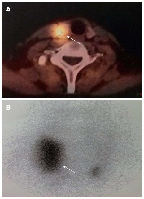
Figure 4 Radio-isotope scan reveals soft tissue masses (arrows) posterior to the thyroid gland which displayed abnormal uptake of 99mTc-MIBI.
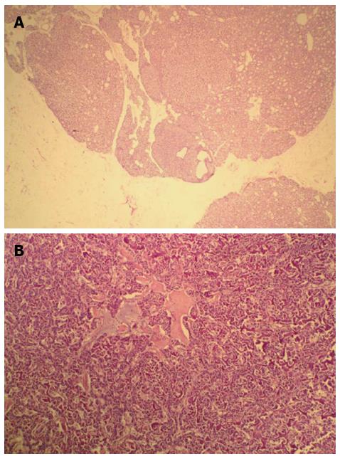
Figure 5 Histological and Pathological analysis of the case (hematoxylin and eosin staining, × 100).
A: Histological analysis shows chief cell hyperplasia of parathyroid gland; B: Pathological analysis shows that tumor cells had acidophilic cytoplasm and round nucleoli which were uniform in size and shape, arranged in tubular, organoid and gyriform patterns.
- Citation: Lu YY, Zhu F, Jing DD, Wu XN, Lu LG, Zhou GQ, Wang XP. Multiple endocrine neoplasia type 1 with upper gastrointestinal hemorrhage and perforation: A case report and review. World J Gastroenterol 2013; 19(8): 1322-1326
- URL: https://www.wjgnet.com/1007-9327/full/v19/i8/1322.htm
- DOI: https://dx.doi.org/10.3748/wjg.v19.i8.1322


