Copyright
©2013 Baishideng Publishing Group Co.
World J Gastroenterol. Feb 21, 2013; 19(7): 1135-1139
Published online Feb 21, 2013. doi: 10.3748/wjg.v19.i7.1135
Published online Feb 21, 2013. doi: 10.3748/wjg.v19.i7.1135
Figure 1 Computed tomography.
A: Abdomen and pelvis showing massive hepatosplenomegaly at initial presentation; B: Chest at initial presentation demonstrating extensive bilateral emphysematous changes; C: Abdomen revealing pockets of free air consistent with perforation. No evidence of pneumatosis is seen on computed tomography scan. A: Anterior; P: Posterior; R: Right; L: Left.
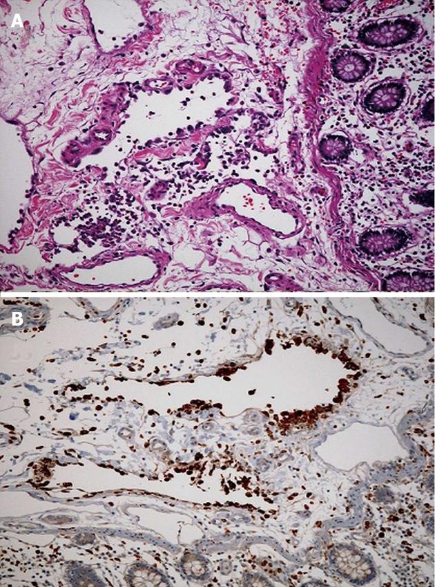
Figure 2 Pathology of the resected tissue revealed portions of large bowel with submucosal and mucosal hemorrhage and submucosa with irregular spaces lined with histiocytes (CD68 positive and CD31 negative) consistent with pneumatosis cystoides intestinalis.
A: Cecum specimen stained with hematoxylin and eosin demonstrates partially endotheliolized cystic spaces in the submucosa lined by eosinophilic cells characteristic of pneumatosis intestinalis; B: Immunohistochemical staining for CD 68 which is found in cytoplasmic granules of cells within the monocyte/macrophage lineage confirms presents of histiocytes lining cystic spaces.
Figure 3 Computed tomography abdomen with contrast demonstrating marked reduction in spleen size and moderate reduction in hepatomegaly after 5 mo of steroid treatment.
A: Anterior; P: Posterior; R: Right; L: Left.
- Citation: Rahim H, Khan M, Hudgins J, Lee K, Du L, Amorosa L. Gastrointestinal sarcoidosis associated with pneumatosis cystoides intestinalis. World J Gastroenterol 2013; 19(7): 1135-1139
- URL: https://www.wjgnet.com/1007-9327/full/v19/i7/1135.htm
- DOI: https://dx.doi.org/10.3748/wjg.v19.i7.1135


