Copyright
©2013 Baishideng Publishing Group Co.
World J Gastroenterol. Dec 14, 2013; 19(46): 8630-8637
Published online Dec 14, 2013. doi: 10.3748/wjg.v19.i46.8630
Published online Dec 14, 2013. doi: 10.3748/wjg.v19.i46.8630
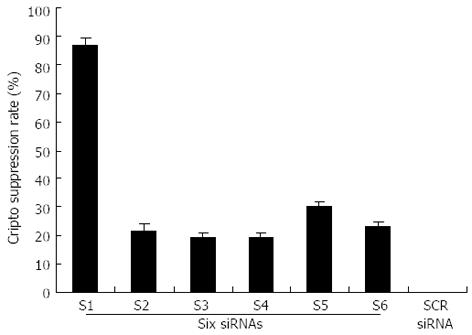
Figure 1 Suppression of cripto mRNA by six siRNA transfection in LS-174T cells.
The colon cancer cell line LS-174T was transfected with 100 nmol/L of six siRNA after 48-h incubation. Real-time polymerase chain reaction quantification of the relative amount of cripto transcript was performed using the cripto primers and probe set as described in “Materials and Methods,” using glyceraldehyde-3-phosphate dehydrogenase (GAPDH) as internal standard. All data are presented as the mean ± SD of three independent experiments; the level of cripto mRNA relative to GAPDH in untreated cells maintained under identical experimental conditions was taken as 100%.
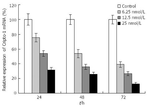
Figure 2 Effects of S1 siRNA on cripto mRNA of colon cancer LS-174T cells.
The LS-174T colon cancer cell line was transfected with different dose of S1 siRNA for 24, 48, 72 h. Cripto mRNA was evaluated by real-time reverse transcription-polymerase chain reaction as described in “Materials and Methods,” using GAPDH as internal standard. All data are presented as the mean ± SD of three independent experiments; the level of cripto mRNA relative to GAPDH in untreated cells maintained under identical experimental conditions was taken as 100%.
Figure 3 Effects of S1 siRNA on cell proliferation and anchorage-independent growth of colon cancer LS-174T cells.
A: S1 siRNA treatment decreases cell proliferation. Cells plated in 96-well plates were grown in their respective media for 48 h after the addition of siRNA. Cell proliferation was examined by MTT assay; B: S1 siRNA treatment reduces anchorage-independent growth of LS-174T cells. The anchorage-independent growth was evaluated by colony formation in soft agar.
Figure 4 Effects of S1 siRNA on migration and invasion of colon cancer LS-174T cells.
The LS-174T colon cancer cell line was used in this experiment. Cell migration was assessed in BioCoat control cell culture chambers. Transwell migration and invasion assays were performed using LS-174T cells cultured in 12-well plates containing either Matrigel-coated inserts (invasion assays) or 8 μm pore Biocoat® control inserts (migration assays), according to the manufacturer’s instructions. Control, scrambled siRNA-, and S1 siRNA-treated cells were added to control and Matrigel chambers. A: The percentage of invasion was expressed as the ratio of mean cell number from invasion chamber to mean cell number from control chamber according to the manufacturer’s recommendation; B: The percentage of migration was expressed as the ratio of mean cell number in control inserts containing siRNA-treated cells to mean cell number in control inserts containing untreated control cells. Control cells were given a value of 100%.
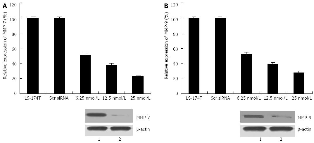
Figure 5 Effects of S1 treatment on matrix metalloproteinase-7 and -9 expression of colon cancer line LS-174T.
After cancer cells were treated with S1 siRNA for different times, cells were harvested, and total RNA and proteins were extracted. Real-time reverse transcription-polymerase chain reaction and western blotting were performed to detect mRNA and protein levels, respectively, of matrix metalloproteinase (MMP)-7 and -9. A: Expression level of mRNA and protein of MMP-7 (2 h); B: Expression level of mRNA and protein of MMP-9 (72 h). 1: Scr siRNA; 2: S1(25 nmol/L).
-
Citation: Jiang PC, Zhu L, Fan Y, Zhao HL. Clinicopathological and biological significance of
cripto overexpression in human colon cancer. World J Gastroenterol 2013; 19(46): 8630-8637 - URL: https://www.wjgnet.com/1007-9327/full/v19/i46/8630.htm
- DOI: https://dx.doi.org/10.3748/wjg.v19.i46.8630


