Copyright
©2013 Baishideng Publishing Group Co.
World J Gastroenterol. Dec 7, 2013; 19(45): 8453-8458
Published online Dec 7, 2013. doi: 10.3748/wjg.v19.i45.8453
Published online Dec 7, 2013. doi: 10.3748/wjg.v19.i45.8453
Figure 1 Computed tomography.
A: Distal pancreatic tumor; B: Hydrops adjacent to the pancreatic stump; C: A pigtail catheter (percutaneous catheter drainage tube 1) was placed into the left anterior pararenal space under computed tomography (CT) guidance; D: Abdominal contrast-enhanced CT revealed extravasation of contrast medium adjacent to the pancreatic stump.
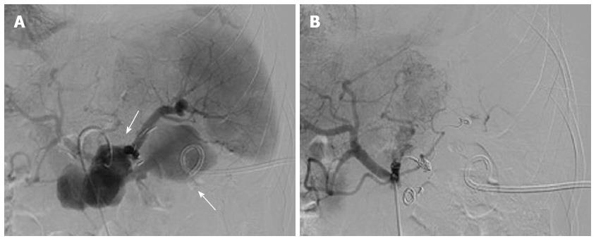
Figure 2 Celiac trunk angiography.
A: Celiac trunk angiography revealed pseudoaneurysm of the splenic artery; B: Celiac trunk angiography revealed no extravasation of contrast medium after embolization of the splenic artery with microcoils.
Figure 3 Examination.
A: Contrast-enhanced abdominal computed tomography revealed a peripancreatic effusion; B: Multiple focal ischemic lesions of the spleen; C: A pigtail catheter (percutaneous catheter drainage tube 2) was placed to drain the peripancreatic fluid collection; D: Absorption of the peripancreatic effusion.
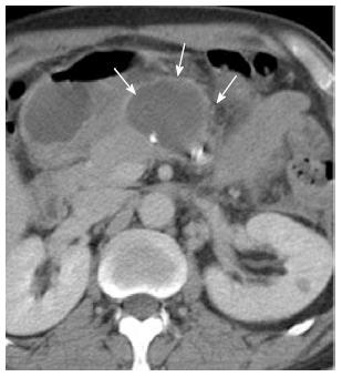
Figure 4 Encapsulated fluid collection anterior to the pancreatic stump.
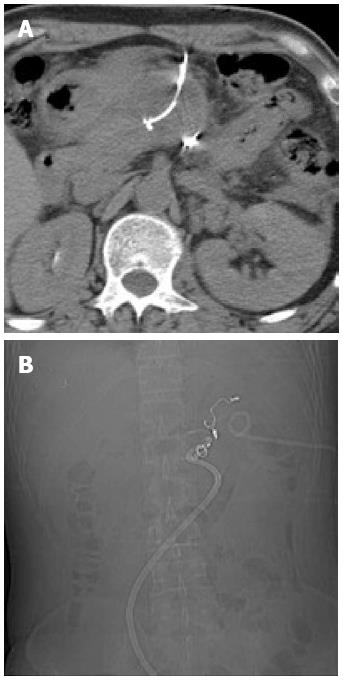
Figure 5 A pigtail catheter (percutaneous catheter drainage tube 3) was placed anterior to the pancreatic stump to drain the effusion.
A: Percutaneous catheter drainage (PCD) tube 3; B: PCD tubes 1 and 3.
- Citation: Zhu YP, Ni JJ, Chen RB, Matro E, Xu XW, Li B, Hu HJ, Mou YP. Successful interventional radiological management of postoperative complications of laparoscopic distal pancreatectomy. World J Gastroenterol 2013; 19(45): 8453-8458
- URL: https://www.wjgnet.com/1007-9327/full/v19/i45/8453.htm
- DOI: https://dx.doi.org/10.3748/wjg.v19.i45.8453


