Copyright
©2013 Baishideng Publishing Group Co.
World J Gastroenterol. Nov 28, 2013; 19(44): 8151-8155
Published online Nov 28, 2013. doi: 10.3748/wjg.v19.i44.8151
Published online Nov 28, 2013. doi: 10.3748/wjg.v19.i44.8151
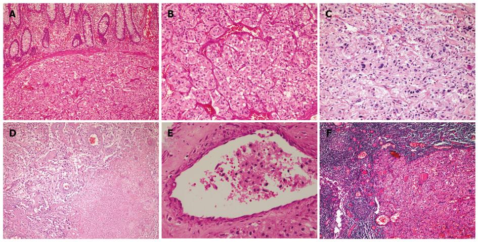
Figure 1 Histological features.
A-B: The tumor was composed of sheets or organoid nests of large polygonal cells surrounded by a rich network of delicate arborizing vasculature, generating a characteristic “zellballen” (A: HE, × 100; B: HE, × 400); C: Focal nuclear pleomorphism (HE, × 400); D: Confluent tumor necrosis (HE, × 100); E: Vascular invasion (HE, × 400); F: Metastases of lymph nodes (HE, × 100).
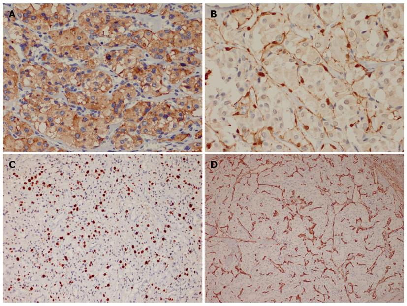
Figure 2 Immunohistochemistry.
A: The tumor cells exhibited diffuse and strong expression of chromogranin A; B: S100 protein highlighted the presence of slender sustentacular cells located at the periphery of the tumor nests; C: The Ki67 index was approximately 20%; D: CD34 outlined the rich vascular network.
- Citation: Yu L, Wang J. Malignant paraganglioma of the rectum: The first case report and a review of the literature. World J Gastroenterol 2013; 19(44): 8151-8155
- URL: https://www.wjgnet.com/1007-9327/full/v19/i44/8151.htm
- DOI: https://dx.doi.org/10.3748/wjg.v19.i44.8151


