Copyright
©2013 Baishideng Publishing Group Co.
World J Gastroenterol. Oct 28, 2013; 19(40): 6931-6933
Published online Oct 28, 2013. doi: 10.3748/wjg.v19.i40.6931
Published online Oct 28, 2013. doi: 10.3748/wjg.v19.i40.6931
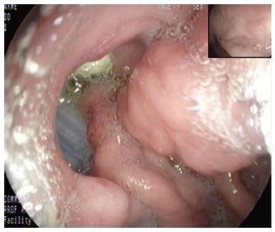
Figure 1 Appearance of a large defect in the posterior wall of the stomach with the post-operative drains seen through it.
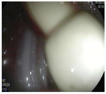
Figure 2 A closer look at the drains through the defect.
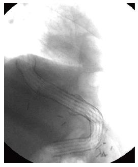
Figure 3 Two co-axially inserted partially covered self-expandable esophageal metal stents extending from the esophagus to the duodenum.
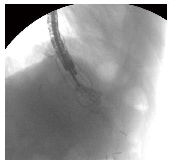
Figure 4 Self expandable metal stent being removed from the distal aspect with a rat-tooth forceps.
- Citation: Almadi MA, Aljebreen AM, Bamihriz F. Resolution of an esophageal leak and posterior gastric wall necrosis with esophageal self-expandable metal stents. World J Gastroenterol 2013; 19(40): 6931-6933
- URL: https://www.wjgnet.com/1007-9327/full/v19/i40/6931.htm
- DOI: https://dx.doi.org/10.3748/wjg.v19.i40.6931


