Copyright
©2013 Baishideng Publishing Group Co.
World J Gastroenterol. Oct 28, 2013; 19(40): 6928-6930
Published online Oct 28, 2013. doi: 10.3748/wjg.v19.i40.6928
Published online Oct 28, 2013. doi: 10.3748/wjg.v19.i40.6928
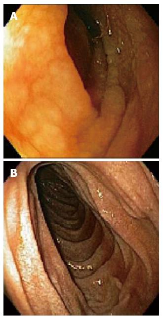
Figure 1 Scalloped mucosa (A) and normal duodenum (B) (endoscopy).
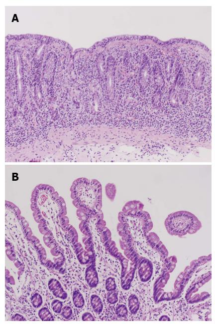
Figure 2 Complete duodenal villous blunting (A) and villous regeneration (B) (hematoxylin and eosin, × 400).
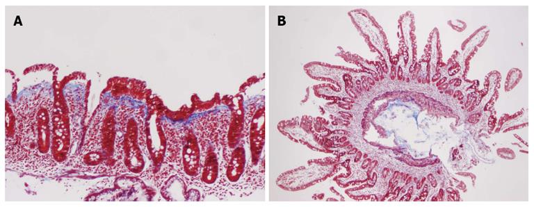
Figure 3 Thickened collagen table (A) and normal histology (B) (trichrome, × 400).
- Citation: Nielsen JA, Steephen A, Lewin M. Angiotensin-II inhibitor (olmesartan)-induced collagenous sprue with resolution following discontinuation of drug. World J Gastroenterol 2013; 19(40): 6928-6930
- URL: https://www.wjgnet.com/1007-9327/full/v19/i40/6928.htm
- DOI: https://dx.doi.org/10.3748/wjg.v19.i40.6928


