Copyright
©2013 Baishideng Publishing Group Co.
World J Gastroenterol. Oct 28, 2013; 19(40): 6869-6875
Published online Oct 28, 2013. doi: 10.3748/wjg.v19.i40.6869
Published online Oct 28, 2013. doi: 10.3748/wjg.v19.i40.6869
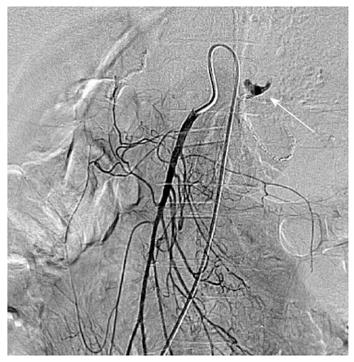
Figure 1 A 35-year-old man with massive upper gastrointestinal bleeding after gastrectomy of gastric carcinoma.
Selective superior mesenteric arteriography showed active arterial contrast extravasation (white arrow) from the jejunal artery.
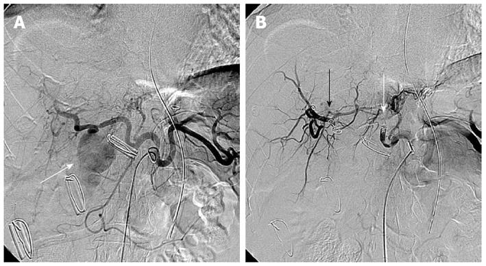
Figure 2 A 56-year-old man presented with massive bleeding from surgical drains after choledocholithotomy due to common bile duct stone.
A: Selective celiac arteriography showed a pseudoaneurysm (arrow) arising from the right hepatic artery; B: Selective celiac arteriography after embolization with microcoils and gelfoam demonstrated disappearance of the pseudoaneurysm. Embolic agents were inserted proximally (white arrow) and distally (black arrow) to the origin of the pseudoaneurysm.
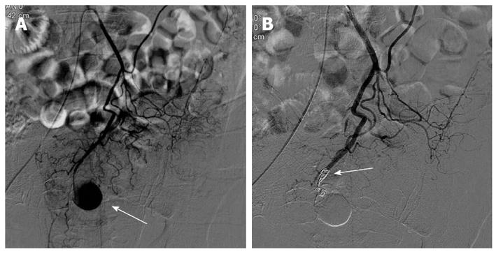
Figure 3 A 61-year-old man presented with massive bleeding from surgical drains 3 d after excision of the intra-abdominal abscess.
A: Selective inferior mesenteric arteriography shows a pseudoaneurysm (arrow) arising from the superior rectal artery; B: Selective inferior mesenteric arteriography after embolization with microcoils demonstrated complete occlusion (arrow) of the distal end of the superior rectal artery.
- Citation: Zhou CG, Shi HB, Liu S, Yang ZQ, Zhao LB, Xia JG, Zhou WZ, Li LS. Transarterial embolization for massive gastrointestinal hemorrhage following abdominal surgery. World J Gastroenterol 2013; 19(40): 6869-6875
- URL: https://www.wjgnet.com/1007-9327/full/v19/i40/6869.htm
- DOI: https://dx.doi.org/10.3748/wjg.v19.i40.6869


