Copyright
©2013 Baishideng Publishing Group Co.
World J Gastroenterol. Oct 28, 2013; 19(40): 6825-6833
Published online Oct 28, 2013. doi: 10.3748/wjg.v19.i40.6825
Published online Oct 28, 2013. doi: 10.3748/wjg.v19.i40.6825
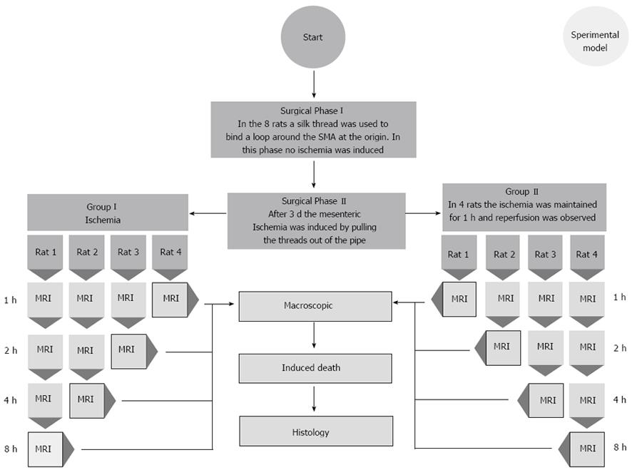
Figure 1 Flow chart.
Flow chart shows our procedure to study the 2 different groups of rats. MRI: Magnetic resonance imaging; SMA: Superior mesenteric artery.
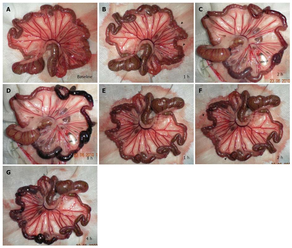
Figure 2 Macroscopic appearance.
A-D: Group I; at baseline evaluation, the bowel loops presented an average diameter of 1.5 mm, uniform serosa and rose-colored mesentery in all segments (A). Pale mesentery and spasm of the jejunal loops (arrowheads) were evident 1 h after acute superior mesenteric artery occlusion (B). Spastic reflex ileus was replaced by definite hypotonic reflex ileus 2 h after acute ischemia (C). Eight hours from acute superior mesenteric artery occlusion, a chromatic change to charcoal black was extended to a large ileal segment (white stars) (D); E-G: Group II; vascular congestion of mesenteric vessels was evident 1 h after reperfusion (E). After 2 h from reperfusion, damaged areas underwent chromatic change from pink to dark red (arrowheads) (F), and became charcoal black after 4 h (stars) (G).
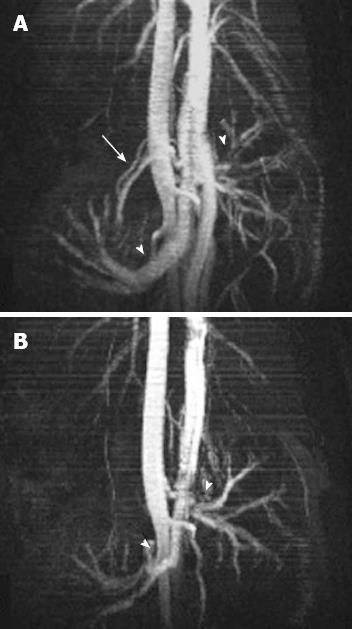
Figure 3 Magnetic resonance imaging angiography.
Maximum intensity projection images through origins of renal arteries (arrowheads) and acute superior mesenteric artery (SMA) (arrow) before (A), and after SMA occlusion (B) (FLASH 2D-TOF), no flow is observed in SMA (B).
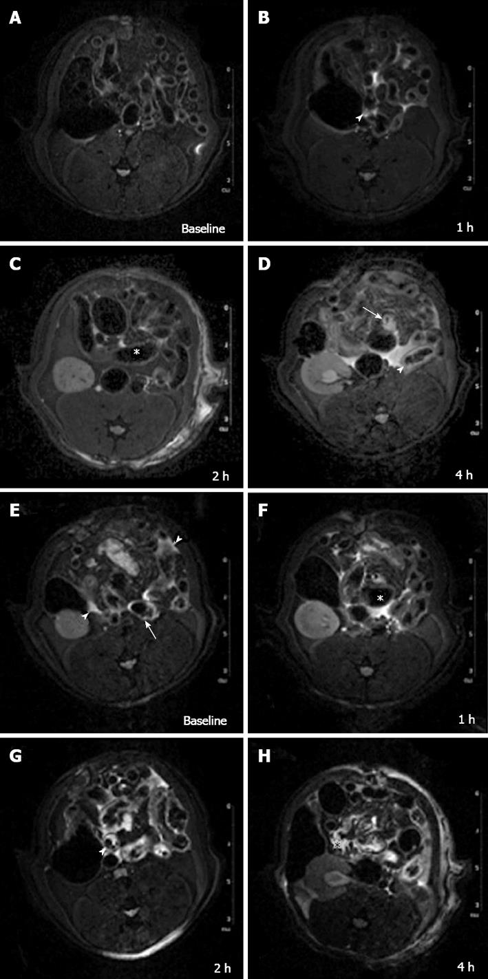
Figure 4 7 Tesla-magnetic resonance imaging appearance.
A-D: Group I; at baseline evaluation no free gas within the abdominal cavity or signs of visceral or mesentery irritation were found on T2-W sequences (A). One hour after acute superior mesenteric artery (SMA) ligation, T2-W sequences showed a thin collection of fluid between the loops (arrowhead) (B). After 2 h dilation of numerous bowel loops [(hypotonic reflex ileus (HRI)] with reduced wall thickness were evident (star) (C). Four hours from SMA ligation HRI was followed by paralytic ileus (PI) (gas-liquid stasis) (arrow); also bowel wall hyper-intense signal was evident in some loops (arrowhead) (D). E-H: Group II; An early hyper-intense signal of bowel wall in some loops (arrow) and a thin layer of peritoneal fluid (arrowheads) were detected 1 h after reperfusion (E). After 2 h, a small amount of peritoneal free fluid, bowel wall thickening, hyper-intense signal of some loops and HRI (star) were observed (F). At 4 h, a paralytic ileus (arrowhead) (G) with a larger amount of peritoneal fluid appeared and a mild mesenteric engorgement (star) became evident beyond a hyper-intense signal of intestinal wall in many loops (H).
- Citation: Saba L, Berritto D, Iacobellis F, Scaglione M, Castaldo S, Cozzolino S, Mazzei MA, Mizio VD, Grassi R. Acute arterial mesenteric ischemia and reperfusion: Macroscopic and MRI findings, preliminary report. World J Gastroenterol 2013; 19(40): 6825-6833
- URL: https://www.wjgnet.com/1007-9327/full/v19/i40/6825.htm
- DOI: https://dx.doi.org/10.3748/wjg.v19.i40.6825


