Copyright
©2013 Baishideng Publishing Group Co.
World J Gastroenterol. Oct 28, 2013; 19(40): 6814-6824
Published online Oct 28, 2013. doi: 10.3748/wjg.v19.i40.6814
Published online Oct 28, 2013. doi: 10.3748/wjg.v19.i40.6814
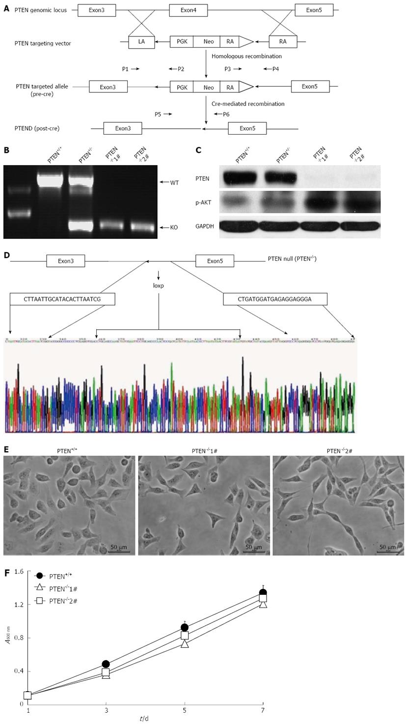
Figure 1 Gene targeting at the PTEN locus.
A: Schematic illustration of the strategy used to inactivate phosphatase and tensin homolog deleted on chromosome 10 (PTEN) by somatic cell gene targeting. The primers (P1, P2, P3, P4, P5, and P6) used for polymerase chain reaction (PCR)-based genotyping are indicated; B: Identification of the desired targeted clones by PCR. Primers P5 and P6 were used to amplify a 1,155-bp product from the wild-type PTEN allele and a 522-bp product from the final knockout allele, respectively; C: Western blotting analysis of PTEN expression; D: Confirmation of PTEN exon 4 deletion in PTEN-deficient cells by sequencing; E: Morphology of PTEN+/+ and PTEN-/- cells. F: Determination of cell viability for PTEN+/+ and PTEN-/- cell lines using the MTT assay. PTEN-/-1# and PTEN-/-2# are two PTEN knockout clones. LA: Left arm; RA: Right arm; GAPDH: Glyceraldehyde-3-phosphate dehydrogenase.
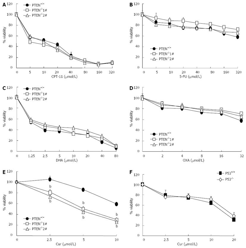
Figure 2 Curcumin shows enhanced cytotoxicity toward PTEN-deficient cells.
A-E: Cytotoxic effects of irinotecan (CPT-11), 5-fluorouracil (5-FU), dihydroartemisinin (DHA), oxaliplatin (OXA) and curcumin (Cur) on cell viability of HCT116 phosphatase and tensin homolog deleted on chromosome 10 (PTEN)+/+ and PTEN-/- cells were determined using the MTT assay; F: Cytotoxic effects of curcumin (Cur) on HCT116 p53+/+ and p53-/- cells ( bP < 0.01 vs control group).
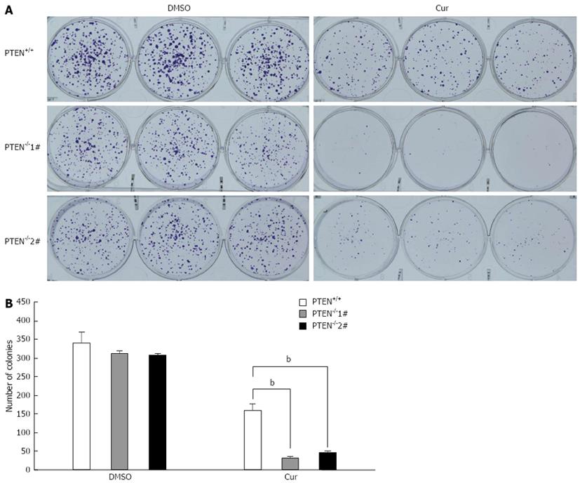
Figure 3 Curcumin has an increased growth-inhibitory effect on PTEN-deficient cells in clonogenic assays.
A: Photographs of clonogenic assay plates; B: Histograms showing clonogenic assay results (bP < 0.01 vs control group). Cur: Curcumin.
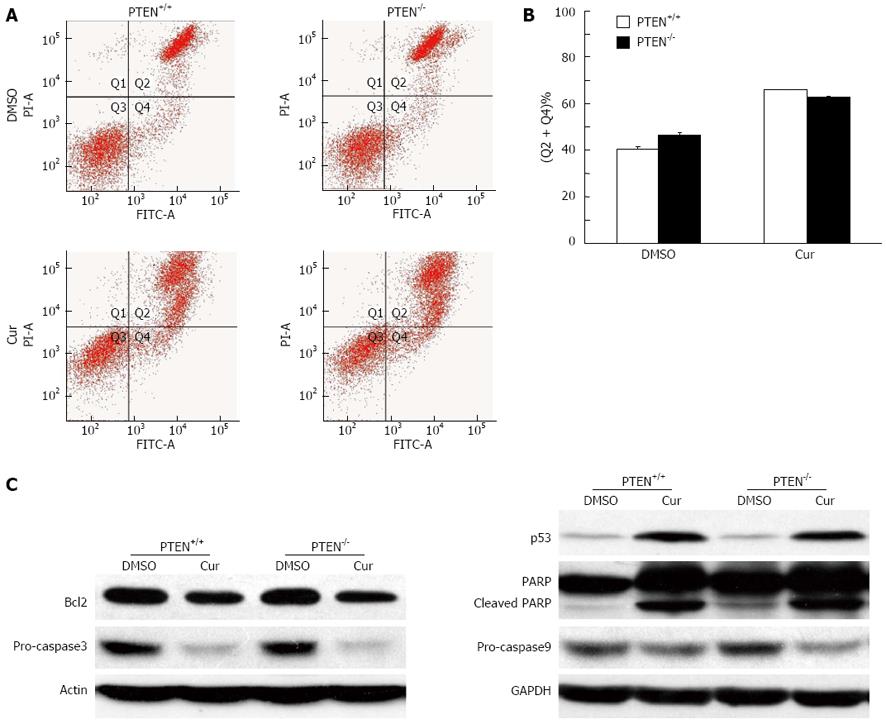
Figure 4 PTEN deficiency does not enhance curcumin-induced apoptosis.
A: Images showing flow cytometric analysis of apoptosis. Apoptosis was analyzed by flow cytometry with the Annexin V-FITC kit; B: Histograms showing apoptosis assay results. Phosphatase and tensin homolog deleted on chromosome 10 (PTEN) deletion did not result in increased apoptosis following curcumin treatment; C: Western blotting analysis. Cell lysates were separated on SDS-PAGE, transferred to polyvinylidene difluoride membranes, and incubated with indicated antibodies. Cur: Curcumin; PI: Propidium iodide; FITC: Fluorescein isothiocyanate; GAPDH: Glyceraldehyde-3-phosphate dehydrogenase.
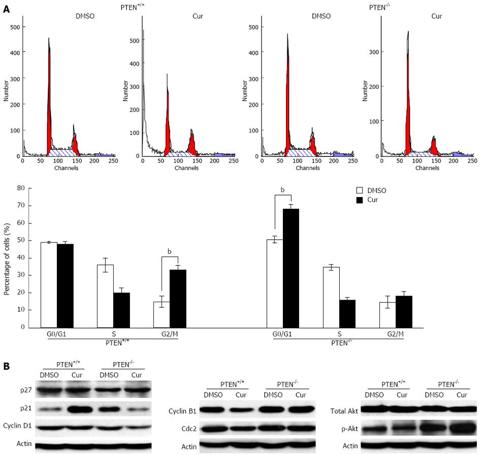
Figure 5 Loss of PTEN alters curcumin-mediated cell cycle arrest.
A: Cell cycle distributions of phosphatase and tensin homolog deleted on chromosome 10 (PTEN)+/+ (left) and PTEN-/- (right) cell samples were analyzed by flow cytometry (bP < 0.01 vs control group); B: Western blotting analysis. Whole cell lysate isolated from PTEN+/+ and PTEN-/- cells were separated on sodium dodecyl sulfate-polyacrylamide gel electrophoresis, transferred to polyvinylidene difluoride membranes, and incubated with the indicated antibodies. Cur: Curcumin.
- Citation: Chen L, Li WF, Wang HX, Zhao HN, Tang JJ, Wu CJ, Lu LT, Liao WQ, Lu XC. Curcumin cytotoxicity is enhanced by PTEN disruption in colorectal cancer cells. World J Gastroenterol 2013; 19(40): 6814-6824
- URL: https://www.wjgnet.com/1007-9327/full/v19/i40/6814.htm
- DOI: https://dx.doi.org/10.3748/wjg.v19.i40.6814


