Copyright
©2013 Baishideng Publishing Group Co.
World J Gastroenterol. Oct 21, 2013; 19(39): 6683-6688
Published online Oct 21, 2013. doi: 10.3748/wjg.v19.i39.6683
Published online Oct 21, 2013. doi: 10.3748/wjg.v19.i39.6683
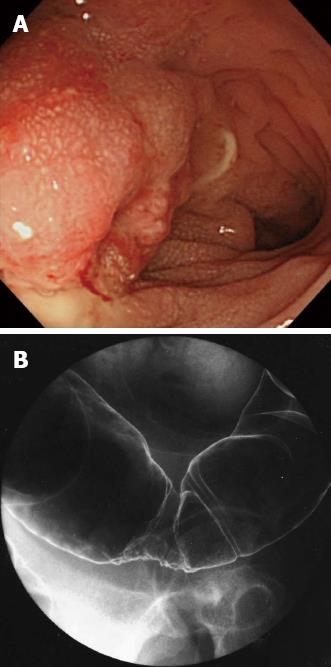
Figure 1 Findings of preoperative examinations.
A: Colonoscopy. B: Barium enema.
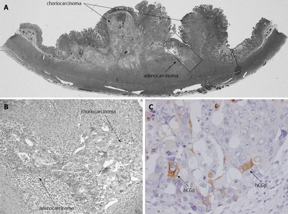
Figure 2 Microscopic findings.
A, B: HE staining shows the co-existence of choriocarcinoma and adenocarcinoma cells (A: loupe, B: × 200); C: The tumor cells were positive for β-human chorionic gonadotropin (hCGβ) (× 400).
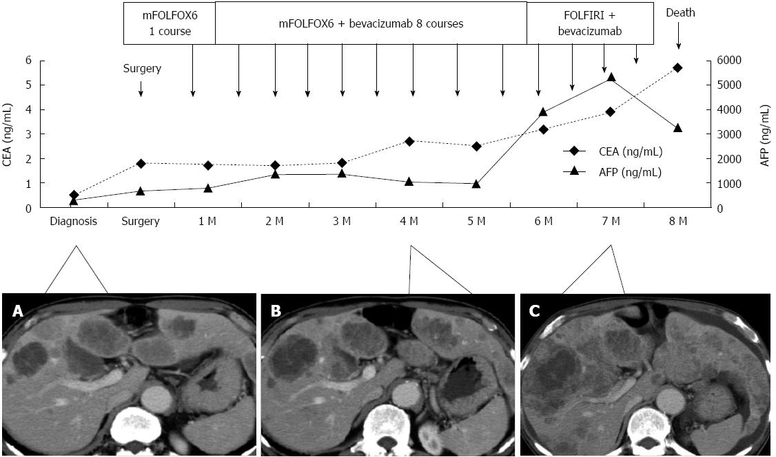
Figure 3 Clinical course of the patient.
A, B: Hepatic metastases showed minimal growth until 4 mo after surgery; at 7 mo after surgery; C: The metastases had markedly enlarged, CEA: Carcinoembryonic antigen; AFP: Alpha-fetoprotein.
- Citation: Maehira H, Shimizu T, Sonoda H, Mekata E, Yamaguchi T, Miyake T, Ishida M, Tani T. A rare case of primary choriocarcinoma in the sigmoid colon. World J Gastroenterol 2013; 19(39): 6683-6688
- URL: https://www.wjgnet.com/1007-9327/full/v19/i39/6683.htm
- DOI: https://dx.doi.org/10.3748/wjg.v19.i39.6683


