Copyright
©2013 Baishideng Publishing Group Co.
World J Gastroenterol. Oct 21, 2013; 19(39): 6651-6655
Published online Oct 21, 2013. doi: 10.3748/wjg.v19.i39.6651
Published online Oct 21, 2013. doi: 10.3748/wjg.v19.i39.6651
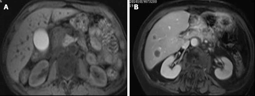
Figure 1 T1 weighted image.
A: T1 weighted image. The margins were not sharp; B: T1 weighted image with contrast. There was obvious enhancement. Two nodules in the right liver demonstrated ring enhancement.
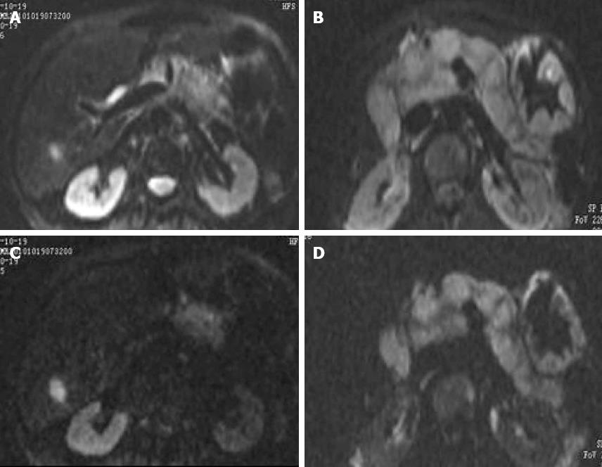
Figure 2 Tumor tissue definition was high, and there was sharp contrast with the surrounding tissue.
A: b = 50 s /mm2; B: b = 400 s /mm2; C: b = 700 s /mm2; D: b = 1100 s /mm2. A high intensity signal was seen.
-
Citation: Hao JG, Wang JP, Gu YL, Lu ML. Importance of
b value in diffusion weighted imaging for the diagnosis of pancreatic cancer. World J Gastroenterol 2013; 19(39): 6651-6655 - URL: https://www.wjgnet.com/1007-9327/full/v19/i39/6651.htm
- DOI: https://dx.doi.org/10.3748/wjg.v19.i39.6651


