Copyright
©2013 Baishideng Publishing Group Co.
World J Gastroenterol. Oct 21, 2013; 19(39): 6637-6644
Published online Oct 21, 2013. doi: 10.3748/wjg.v19.i39.6637
Published online Oct 21, 2013. doi: 10.3748/wjg.v19.i39.6637
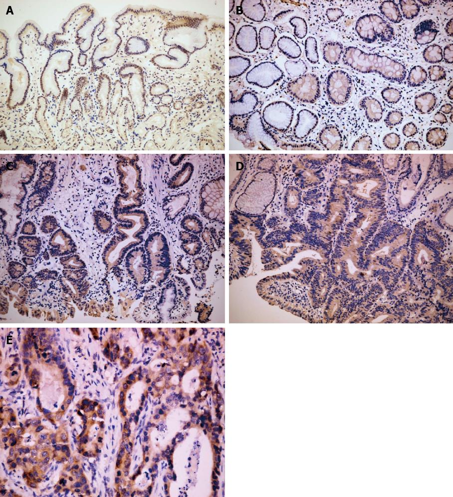
Figure 1 Immunohistochemical staining for Musashi-1 in different gastric tissues.
A: A weak expression of Musashi-1 (Msi-1) was observed in the isthmus of normal gastric glands (× 100); B: The expression of Msi-1 was significantly increased in intestinal metaplastic mucosa (× 100); C: Msi-1 expression showed no significant difference between in low grade intraepithelial neoplasia (× 100); D: high grade intraepithelial neoplasia (× 100); E: The expression of Msi-1 was increased again in the intestinal type gastric cancer (× 400).
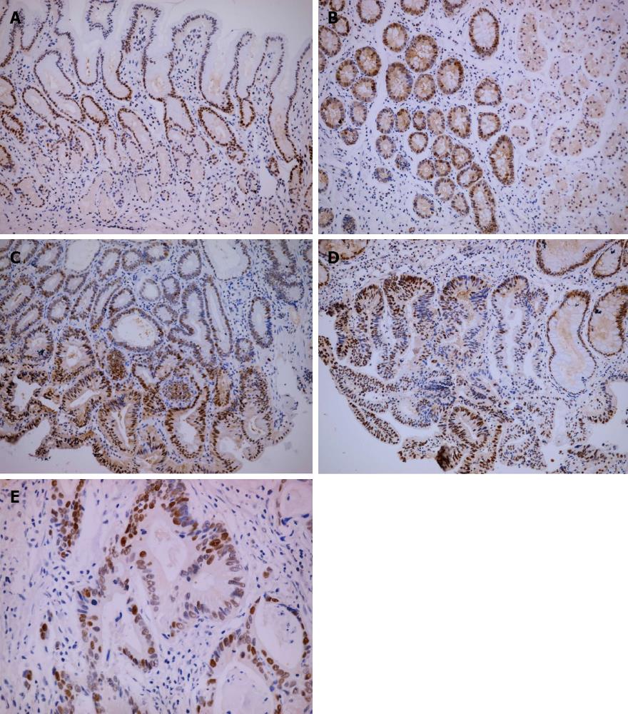
Figure 2 Immunohistochemical staining for proliferating cell nuclear antigen was increased along with the development of gastric carcinogenesis.
A: The staining of proliferating cell nuclear antigen (PCNA) in the isthmus of normal gastric glands (× 100); B: Intestinal metaplastic mucosa (× 100); C: Low grade intraepithelial neoplasia (× 100); D: High grade intraepithelial neoplasia (× 100); E: Strong staining of PCNA in intestinal type gastric cancer (× 400).
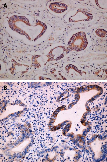
Figure 3 Musashi-1 expresses higher in intestinal type gastric cancer classified as stage II-IV.
A: Type gastric cancer classified as stage II-IV; B: Those classified as stage I (× 400).
- Citation: Kuang RG, Kuang Y, Luo QF, Zhou CJ, Ji R, Wang JW. Expression and significance of Musashi-1 in gastric cancer and precancerous lesions. World J Gastroenterol 2013; 19(39): 6637-6644
- URL: https://www.wjgnet.com/1007-9327/full/v19/i39/6637.htm
- DOI: https://dx.doi.org/10.3748/wjg.v19.i39.6637


