Copyright
©2013 Baishideng Publishing Group Co.
World J Gastroenterol. Oct 7, 2013; 19(37): 6228-6236
Published online Oct 7, 2013. doi: 10.3748/wjg.v19.i37.6228
Published online Oct 7, 2013. doi: 10.3748/wjg.v19.i37.6228
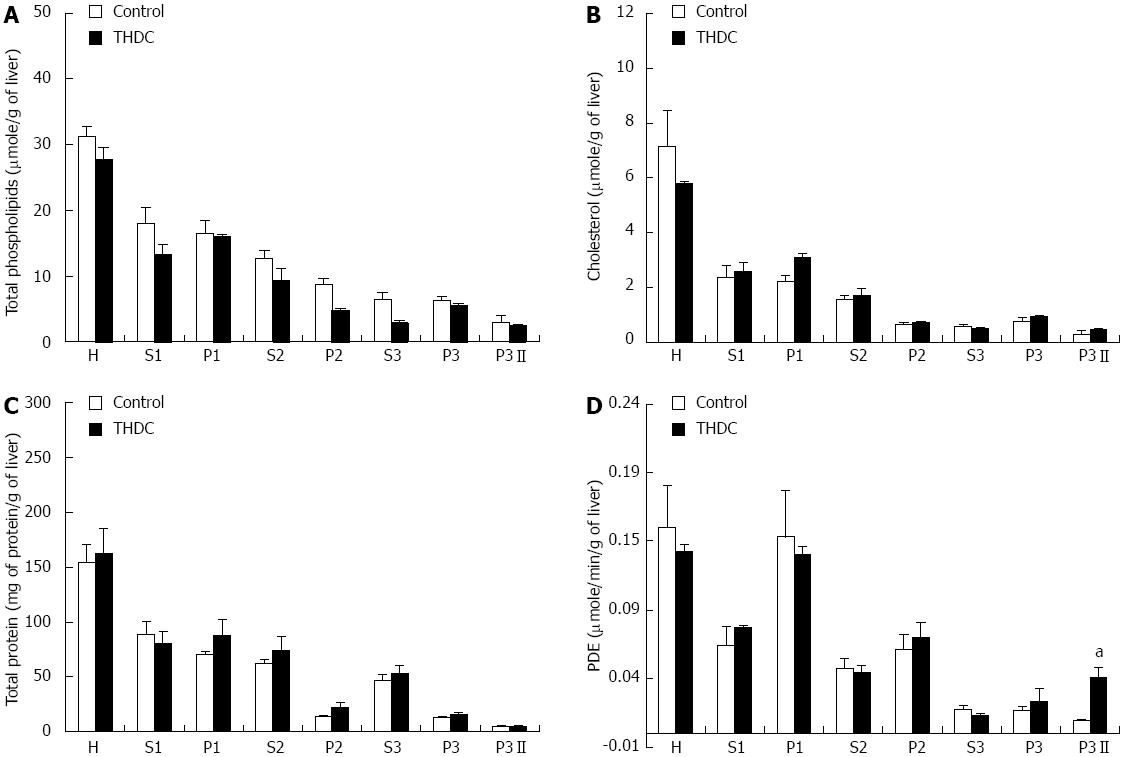
Figure 1 Total phospholipids, cholesterol, protein and phosphodiesterase I activity in subcellular fractions of liver homogenate.
A: Phospholipids; B: Cholesterol; C: Protein; D: Phosphodiesterase I activity (PDE). THDC: Taurohyodeoxycholic acid. Symbols are homogenate (H), supernatant 1 (S1), pellet 1 (P1), supernatant 2 (S2), pellet 2 (P2), supernatant 3 (S3), pellet 3 (P3) and pellet 3 (P3II) diluted with sucrose and KCI. Results are presented as mean ± SE (n = 6). Significant differences from controls were assessed by student’s t-test and are indicated by (aP < 0.05 vs control group).
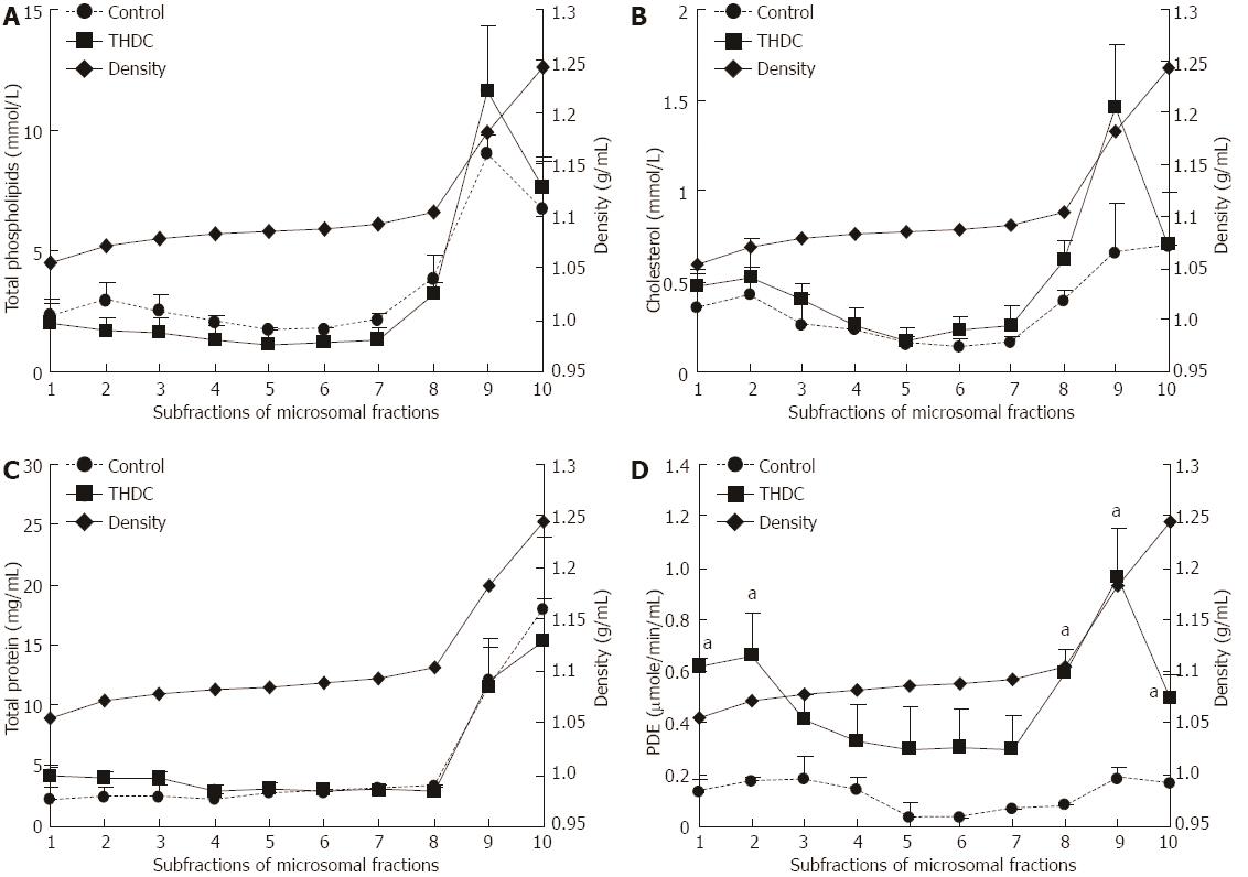
Figure 2 Analysis of the subfractions of the microsomal fraction.
A: Phospholipids; B: Cholesterol; C: Protein; D: Phosphodiesterase I activity (PDE). Livers were removed and the microsomal fraction was prepared and subjected to centrifugation and sub-fractions obtained. The sub-fractions were analyzed for total phospholipids, total cholesterol, protein and PDE activity. Results are presented as mean ± SE (n = 6). THDC: Taurohyodeoxycholic acid. Significant differences from controls were assesses by student’s t test and indicated by aP < 0.05 vs control group.
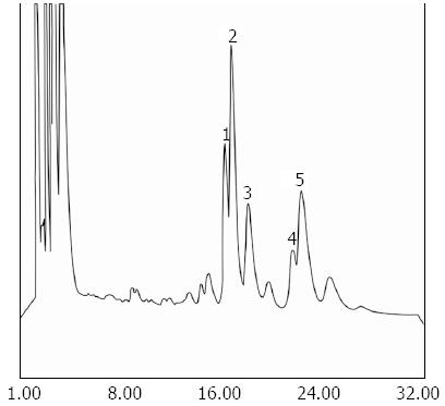
Figure 3 A typical high-performance liquid chromatography chromatogram of a liver homogenate.
Phospholipids were separated by thin layer chromatography (TLC) and the phosphatidylcholine band was scraped off the TLC plate and extracted with 2 × 1 mL methanol [high-performance liquid chromatography (HPLC) grade]. After determination of the phospholipid, 20 nmol was injected onto HPLC for the analysis of the fatty acid composition of the phosphatidylcholine as described in the methods section. Results are presented as mean ± SE (n = 6). A typical chromatogram is presented. Peak 1: Not identified; peak 2: 1-palmitoyl 2-arachidonyl (16:0-20:4) phosphatidylcholine; peak 3: 1-palmitoyl 2-linoleyl (16:0-18:2) phosphatidylcholine; peak 4: 1-palmitoyl 2-oleoyl (16:0-18:1) phosphatidylcholine; peak 5: 1-stearol 2-arachidonyl (18:0-20:4) phosphatidylcholine.
- Citation: Hismiogullari AA, Hismiogullari SE, Rahman K. Isolation and biochemical analysis of vesicles from taurohyodeoxycholic acid-infused isolated perfused rat livers. World J Gastroenterol 2013; 19(37): 6228-6236
- URL: https://www.wjgnet.com/1007-9327/full/v19/i37/6228.htm
- DOI: https://dx.doi.org/10.3748/wjg.v19.i37.6228


