Copyright
©2013 Baishideng Publishing Group Co.
World J Gastroenterol. Sep 28, 2013; 19(36): 6108-6109
Published online Sep 28, 2013. doi: 10.3748/wjg.v19.i36.6108
Published online Sep 28, 2013. doi: 10.3748/wjg.v19.i36.6108
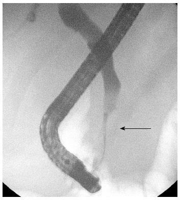
Figure 1 Cholangiogram outlining a distal biliary stricture (arrow).
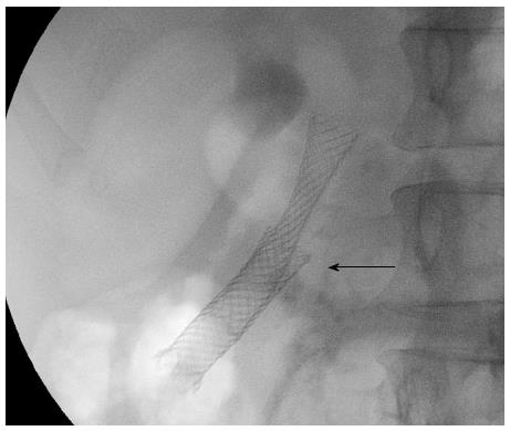
Figure 2 Second covered stent deployed over existing stent (arrow).
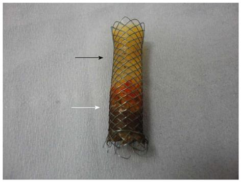
Figure 3 Covered stent after extraction.
The white arrow indicates its duodenal end. There is evidence of damage to the polymer covering of the stent (black arrow).
- Citation: Menon S. Removal of an embedded "covered" biliary stent by the "stent-in-stent" technique. World J Gastroenterol 2013; 19(36): 6108-6109
- URL: https://www.wjgnet.com/1007-9327/full/v19/i36/6108.htm
- DOI: https://dx.doi.org/10.3748/wjg.v19.i36.6108


