Copyright
©2013 Baishideng Publishing Group Co.
World J Gastroenterol. Aug 14, 2013; 19(30): 4992-4999
Published online Aug 14, 2013. doi: 10.3748/wjg.v19.i30.4992
Published online Aug 14, 2013. doi: 10.3748/wjg.v19.i30.4992
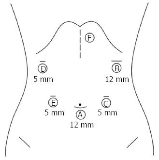
Figure 1 Positions of trocars.
The trocars were inserted into the abdomen in the order A-E. Position F stands for the 4-5 cm midline minilaparotomy incision for reconstruction.
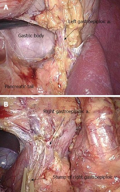
Figure 2 Gastroepiploic artery.
A: Dividing left gastroepiploic artery (arrow); B: Dividing right gastroepiploic vessels (arrow). a: Artery; v: Vein.
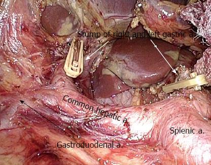
Figure 3 Tracing gastroduodenal artery to locate celiac trunk and its branches (arrows).
a: Artery.
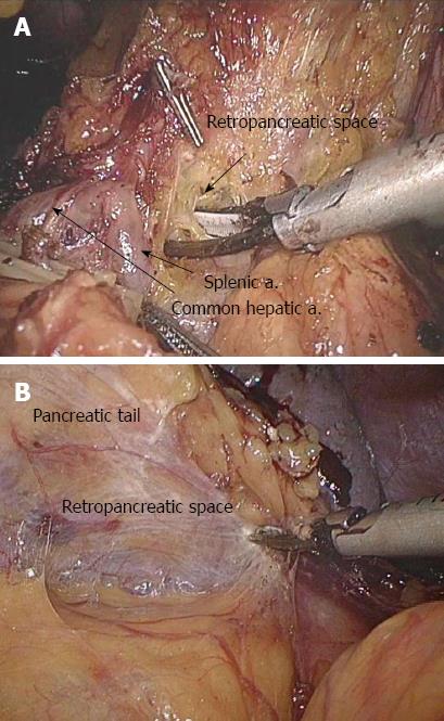
Figure 4 Entering retropancreatic space.
A: Near the superior margin of the pancreas (arrows); B: Near the lower margin of the pancreatic tail. a: Artery.
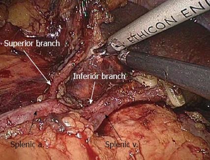
Figure 5 Skeletonizing the branches of the splenic artery (arrows).
a: Artery; v: Vein.
- Citation: Mou TY, Hu YF, Yu J, Liu H, Wang YN, Li GX. Laparoscopic splenic hilum lymph node dissection for advanced proximal gastric cancer: A modified approach for pancreas- and spleen-preserving total gastrectomy. World J Gastroenterol 2013; 19(30): 4992-4999
- URL: https://www.wjgnet.com/1007-9327/full/v19/i30/4992.htm
- DOI: https://dx.doi.org/10.3748/wjg.v19.i30.4992


