Copyright
©2013 Baishideng Publishing Group Co.
World J Gastroenterol. Jul 28, 2013; 19(28): 4630-4632
Published online Jul 28, 2013. doi: 10.3748/wjg.v19.i28.4630
Published online Jul 28, 2013. doi: 10.3748/wjg.v19.i28.4630
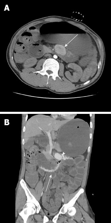
Figure 1 Computed tomography scans.
A: Computed tomography (CT) scanning revealed a superior mesentery artery aneurysm about 3.9 cm in diameter at the jejunal mesentery (white arrow); B: CT scan showing a superior mesentery artery aneurysm (arrow head) with active contrast extravasation into the fourth portion of the duodenum (white arrow).
- Citation: Choo CH, Yen HH. Unusual upper gastrointestinal bleeding: Ruptured superior mesenteric artery aneurysm in rheumatoid arthritis. World J Gastroenterol 2013; 19(28): 4630-4632
- URL: https://www.wjgnet.com/1007-9327/full/v19/i28/4630.htm
- DOI: https://dx.doi.org/10.3748/wjg.v19.i28.4630


