Copyright
©2013 Baishideng Publishing Group Co.
World J Gastroenterol. Jul 28, 2013; 19(28): 4526-4530
Published online Jul 28, 2013. doi: 10.3748/wjg.v19.i28.4526
Published online Jul 28, 2013. doi: 10.3748/wjg.v19.i28.4526
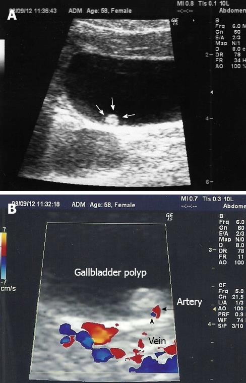
Figure 1 The ultrasonographic image of a 6-mm gallbladder polyp (A) and the same polyp with a feeding artery in Doppler ultrasonography (B).
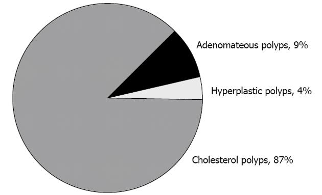
Figure 2 Distribution of pseudopolyp cases.
- Citation: Sarkut P, Kilicturgay S, Ozer A, Ozturk E, Yilmazlar T. Gallbladder polyps: Factors affecting surgical decision. World J Gastroenterol 2013; 19(28): 4526-4530
- URL: https://www.wjgnet.com/1007-9327/full/v19/i28/4526.htm
- DOI: https://dx.doi.org/10.3748/wjg.v19.i28.4526


