Copyright
©2013 Baishideng Publishing Group Co.
World J Gastroenterol. Jun 7, 2013; 19(21): 3300-3308
Published online Jun 7, 2013. doi: 10.3748/wjg.v19.i21.3300
Published online Jun 7, 2013. doi: 10.3748/wjg.v19.i21.3300
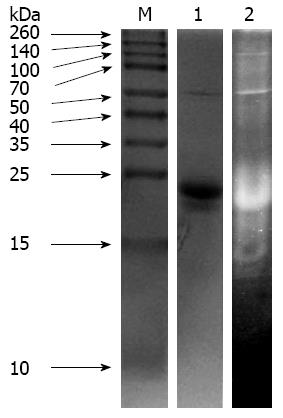
Figure 1 Protein (lane 1) and trypsin inhibitor activity (lane 2) staining of sweet potato protein on polyacrylamide gel electrophoresis gels (12%) with β-mercaptoethanol.
A 10 μL aliquot of staining of sweet potato was loaded in each lane. M: Molecular standards.
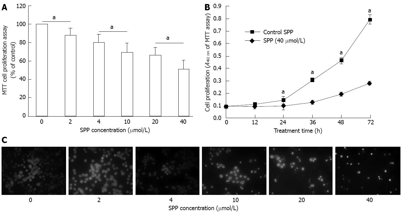
Figure 2 Anti-proliferative effect of sweet potato protein on human colorectal cancer SW480 cells.
A: Effects of various concentrations of sweet potato protein (SPP) on SW480 cell proliferation. We observed dose-dependent inhibition by SPP. aP < 0.05 between groups; B: Effect of various treatment times with 40 μmol/L SPP on SW480 cell proliferation. Time-dependent inhibition was observed. aP < 0.05 vs control; C: Hoechst 33258 nuclear staining. SW480 cells were incubated in the absence or presence of various concentrations of SPP for 48 h, stained with Hoechst 33258 dye, and observed under a fluorescent microscope (magnification, × 400). Images are representative of at least two independent experiments, with similar results.
Figure 3 Effects of various concentrations of sweet potato protein on migration (A) and invasion (B) of SW480 cells in the Transwell assay.
Inhibition of cell migration was observed with 0.8, 8 and 40 μmol/L sweet potato protein (SPP), aP < 0.05 vs control.
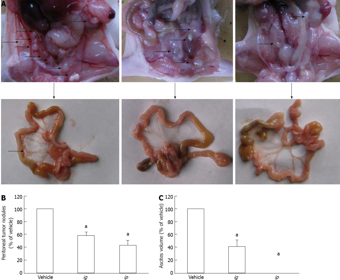
Figure 4 Effect of sweet potato protein on growth of intraperitoneally inoculated human colorectal cancer HCT-8 cells in nude mice.
A: Representative images of growth status of tumor nodules in the peritoneal cavity of nude mice after treatment with intraperitoneally injected or intragastically infused sweet potato protein (SPP); B: Comparison of the number of tumor nodules formed in the peritoneal cavity of nude mice between different treatment groups; C: Volume of bloody ascites generated in the peritoneal cavity of nude mice. Arrows indicate the nodules formed. aP < 0.05 vs vehicle.
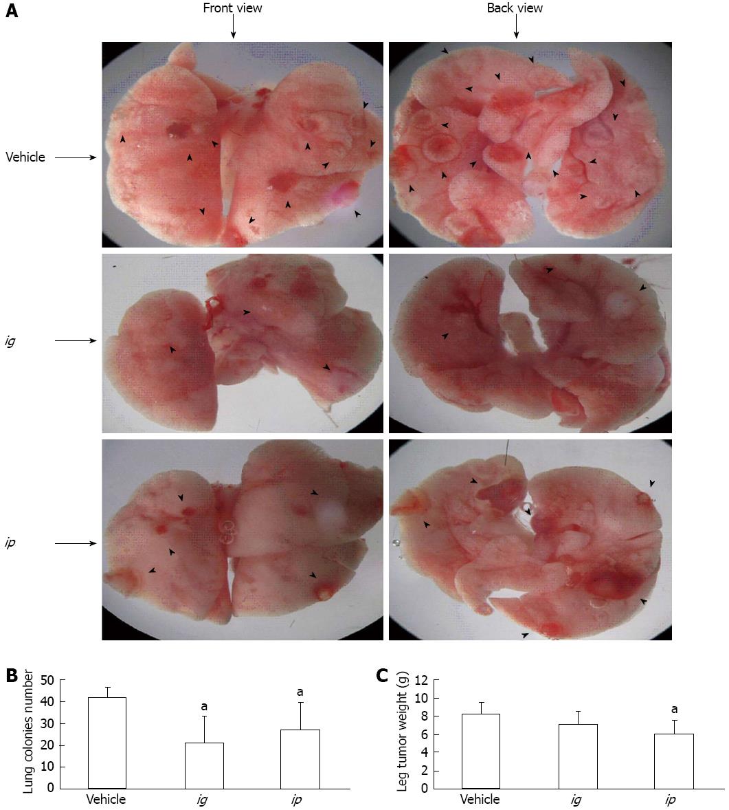
Figure 5 Effect of sweet potato protein on spontaneous metastasis of murine Lewis lung cancer 3LL cells in C57BL/6 mice.
A: Representative images of lungs of mice after inoculation of cancer cells for 25 d; B: Number of spontaneous lung metastatic colonies formed after 25 d of inoculation; C: Weight of subcutaneously inoculated tumor nodules after 25 d. Arrowheads indicate the nodules formed. aP < 0.05 vs vehicle.
- Citation: Li PG, Mu TH, Deng L. Anticancer effects of sweet potato protein on human colorectal cancer cells. World J Gastroenterol 2013; 19(21): 3300-3308
- URL: https://www.wjgnet.com/1007-9327/full/v19/i21/3300.htm
- DOI: https://dx.doi.org/10.3748/wjg.v19.i21.3300


