Copyright
©2013 Baishideng Publishing Group Co.
World J Gastroenterol. Jun 7, 2013; 19(21): 3272-3280
Published online Jun 7, 2013. doi: 10.3748/wjg.v19.i21.3272
Published online Jun 7, 2013. doi: 10.3748/wjg.v19.i21.3272
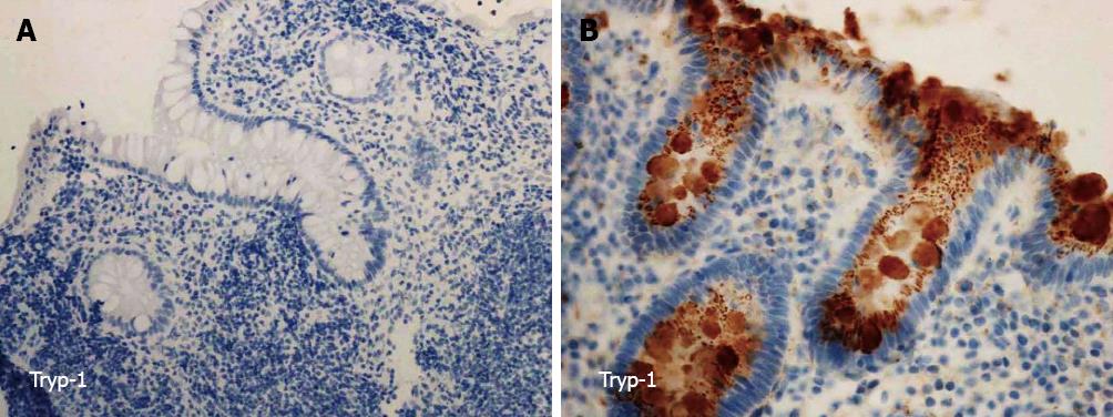
Figure 1 Immunohistochemistry for trypsinogen-1.
A: Lack of immunopositivity for trypsinogen-1 (Tryp-1) in a colonic sample at the diagnosis of a pediatric patient with ulcerative colitis who was operated on two years after diagnosis (× 200); B: Immunopositivity for Tryp-1 in the colonic epithelial cells of a diagnostic tissue sample from a conservatively treated patient with seven years follow-up from the diagnosis (× 400).
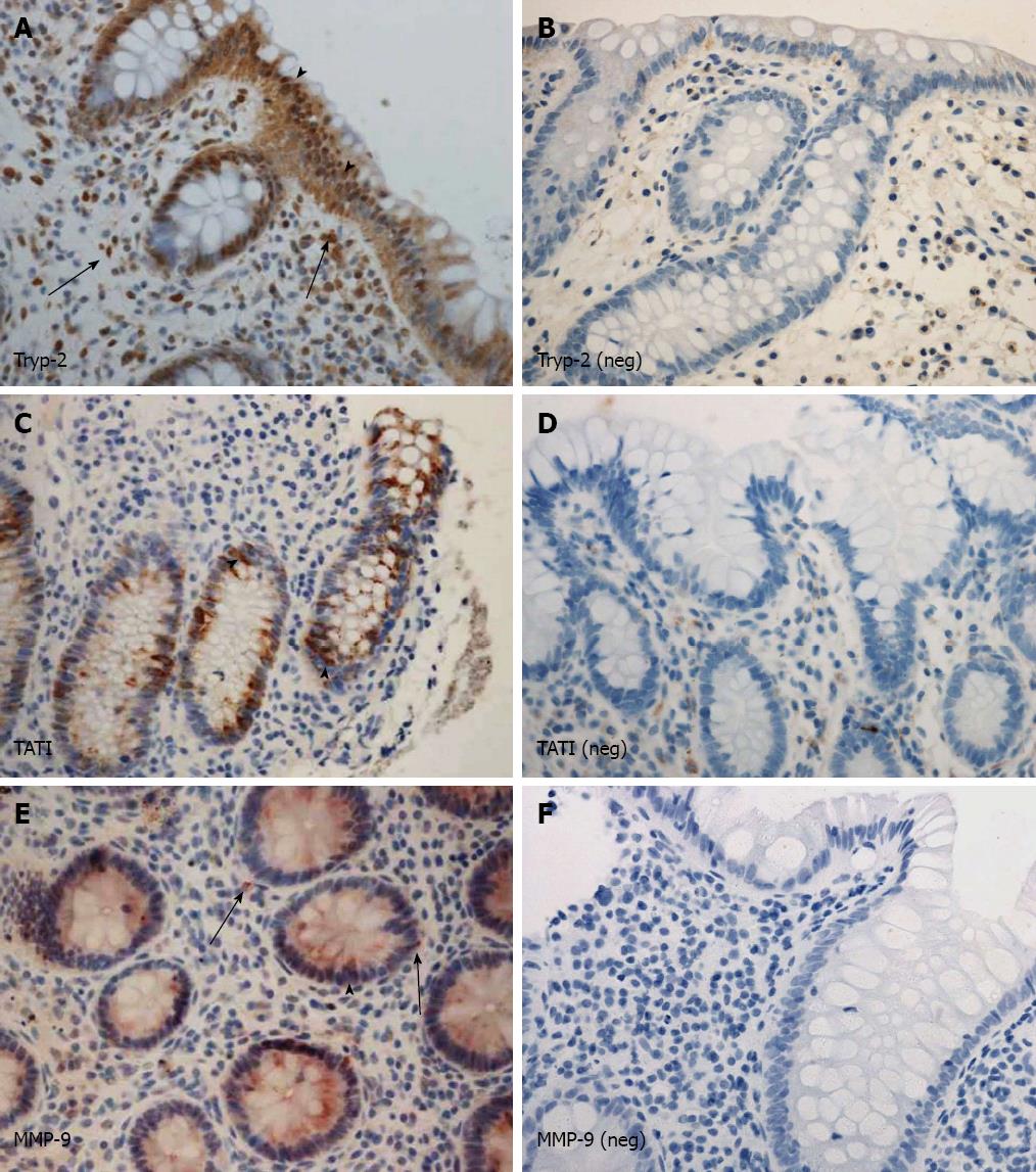
Figure 2 Trypsinogen-2 was stained in the epithelium.
A: Immunopositivity for trypsinogen-2 (Tryp-2); B: Immunonegativity for Tryp-2; C: Immunopositivity for tumor associated trypsin inhibitor (TATI); D: Immunonegativity for TATI; E: Immunopositivity for matrix metalloproteinase-9 (MMP-9); F: Immunonegativity for MMP-9. Arrows (A, E): Immunopositivity in the stromal inflammatory cells; Arrowheads (A, C, E): Immunopositivity in the epithelial cells. A-F, × 400.
- Citation: Piekkala M, Hagström J, Tanskanen M, Rintala R, Haglund C, Kolho KL. Low trypsinogen-1 expression in pediatric ulcerative colitis patients who undergo surgery. World J Gastroenterol 2013; 19(21): 3272-3280
- URL: https://www.wjgnet.com/1007-9327/full/v19/i21/3272.htm
- DOI: https://dx.doi.org/10.3748/wjg.v19.i21.3272


