Copyright
©2013 Baishideng Publishing Group Co.
World J Gastroenterol. Apr 21, 2013; 19(15): 2441-2444
Published online Apr 21, 2013. doi: 10.3748/wjg.v19.i15.2441
Published online Apr 21, 2013. doi: 10.3748/wjg.v19.i15.2441
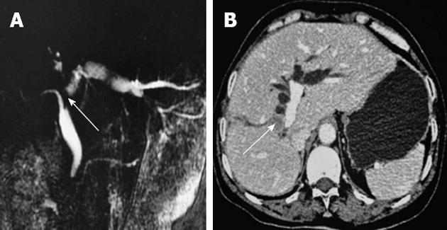
Figure 1 Magnetic resonance cholangiopancreatograph (A)/abdominal enhanced computed tomography (B) showing the hepatic portal soft tissue signal intensity (arrow in B), which is approximately 1.
6 cm in diameter, and the common bile duct proximal locally shows truncated change (arrow in A). The extrahepatic bile duct is widened and the left hepatic intrahepatic bile duct is dilated. Hepatic cirrhosis is present. Images indicate hilar cholangiocarcinoma with intrahepatic bile duct dilatation.
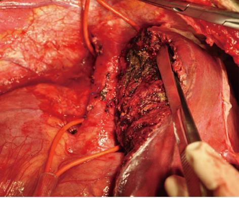
Figure 2 Right hemihepatectomy with caudate process lobectomy and systematic lymphadenectomy of the nodes.
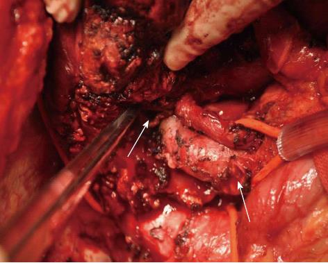
Figure 3 Performing hepatoduodenal ligament lymphadenectomy in hilar cholangiocarcinoma and right hemihepatectomy with caudate process lobectomy.
The left hepatic duct and common bile duct stump are shown (arrows).
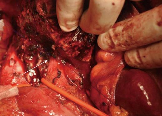
Figure 4 After the radical resection of hilar cholangiocarcinoma, duct-to-duct anastomosis was performed using continuous 5-0 polydioxanone sutures (arrow), with a biliary stent in the bile duct.
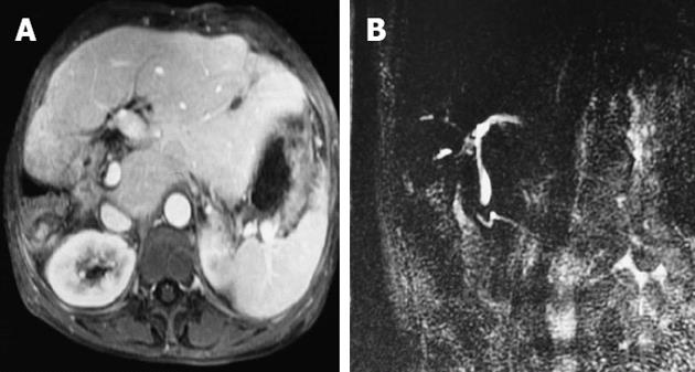
Figure 5 Evidence for tumor recurrence was not found in an magnetic resonance cholangiopancreatograph scan (A, B) 18 mo after operation.
- Citation: Wu WG, Gu J, Dong P, Lu JH, Li ML, Wu XS, Yang JH, Zhang L, Ding QC, Weng H, Ding Q, Liu YB. Duct-to-duct biliary reconstruction after radical resection of Bismuth IIIa hilar cholangiocarcinoma. World J Gastroenterol 2013; 19(15): 2441-2444
- URL: https://www.wjgnet.com/1007-9327/full/v19/i15/2441.htm
- DOI: https://dx.doi.org/10.3748/wjg.v19.i15.2441


