Copyright
©2013 Baishideng Publishing Group Co.
World J Gastroenterol. Apr 7, 2013; 19(13): 2118-2121
Published online Apr 7, 2013. doi: 10.3748/wjg.v19.i13.2118
Published online Apr 7, 2013. doi: 10.3748/wjg.v19.i13.2118
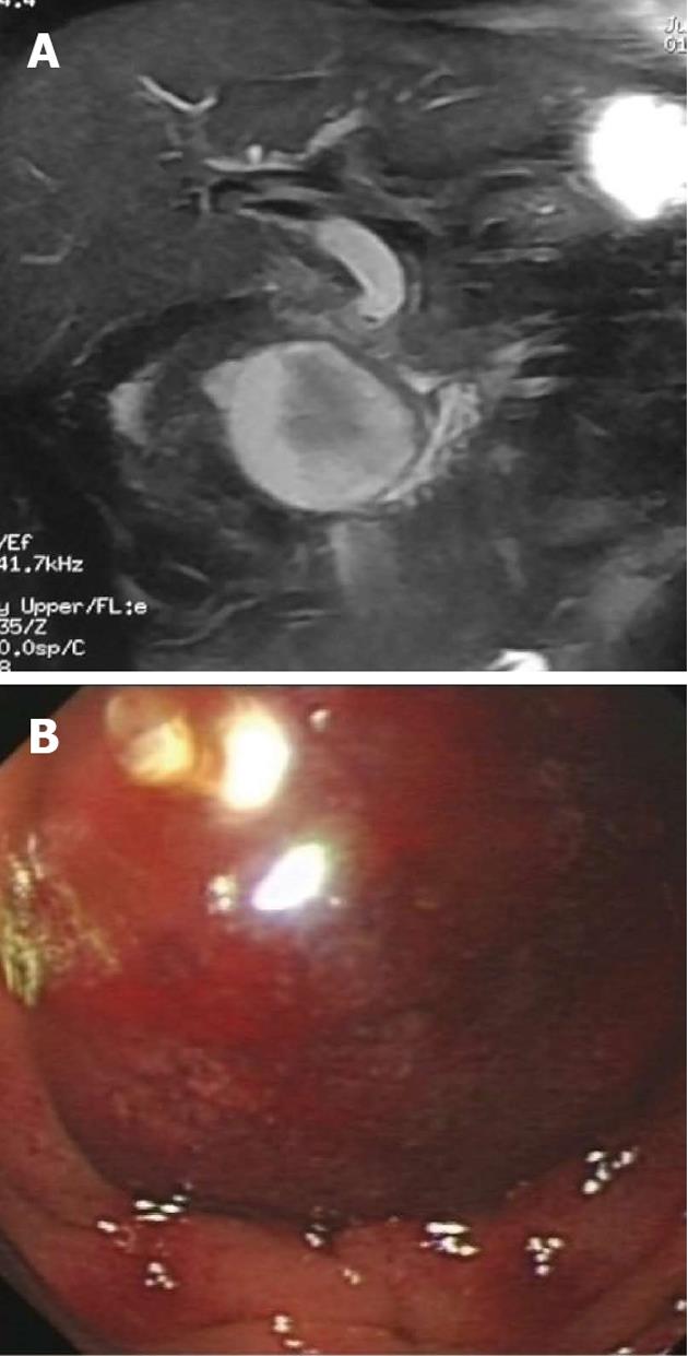
Figure 1 Computed tomography scan and liver function examination of the patient.
A: Computed tomography scan showed a 59 mm × 53 mm intact cyst near the head of the pancreas in the duodenum; B: Duodenoscopy inspection showing a large cystic bulging object in the intestinal wall of the duodenum, obstructing the duodenum.
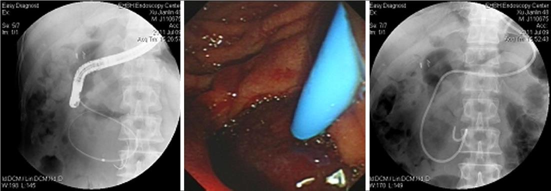
Figure 2 Drainage of the hematoma by the nasal method.
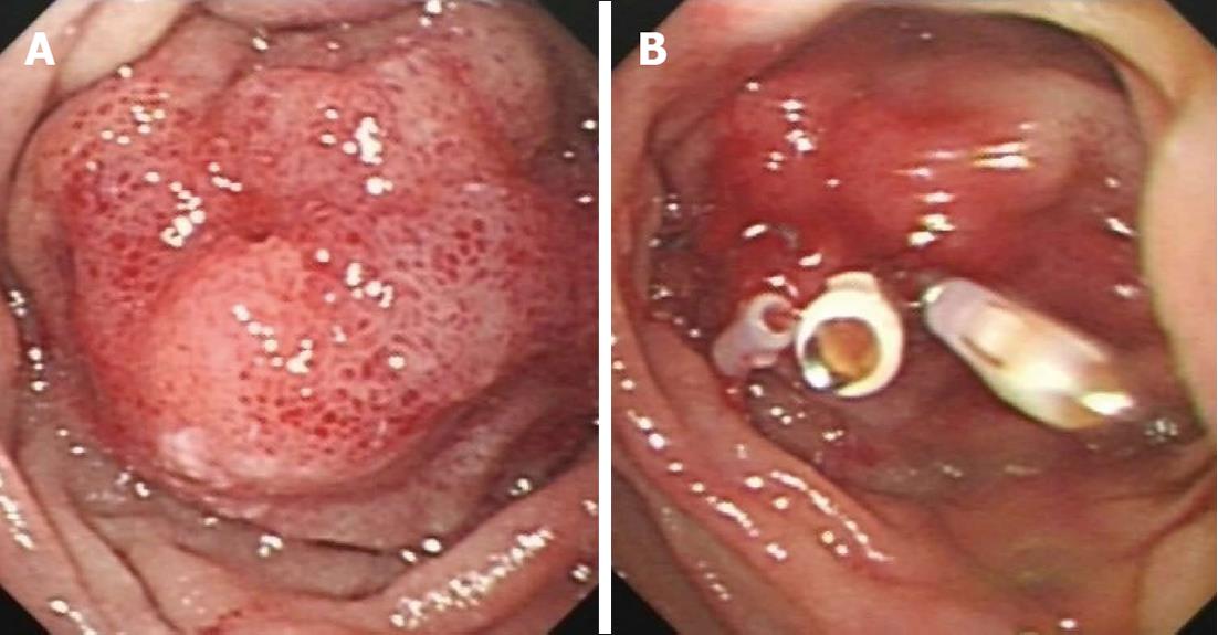
Figure 3 Duodenoscopy examination.
A: Hematoma was greatly decreased in size; B: The puncture area was closed with hemostatic clips.
- Citation: Pan YM, Wang TT, Wu J, Hu B. Endoscopic drainage for duodenal hematoma following endoscopic retrograde cholangiopancreatography: A case report. World J Gastroenterol 2013; 19(13): 2118-2121
- URL: https://www.wjgnet.com/1007-9327/full/v19/i13/2118.htm
- DOI: https://dx.doi.org/10.3748/wjg.v19.i13.2118


