Copyright
©2013 Baishideng Publishing Group Co.
World J Gastroenterol. Apr 7, 2013; 19(13): 2114-2117
Published online Apr 7, 2013. doi: 10.3748/wjg.v19.i13.2114
Published online Apr 7, 2013. doi: 10.3748/wjg.v19.i13.2114
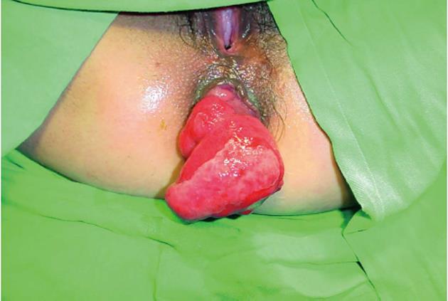
Figure 1 In situ prolapsed angioleiomyoma of the rectum.
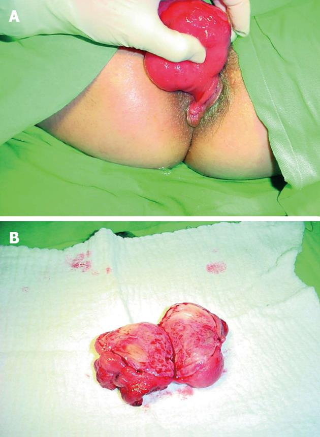
Figure 2 Cross-section of the lobular structure of the tumor showed a visible capsule and a partially bleeding parenchyma.
A: Pedunculated tumor; B: Cross-section of the lobular structure tumor with visible capsule and partially bleeding parenchyma.
Figure 3 Histopathological examination of a combined capillary and venous angioleiomyoma of the rectum.
A: Hematoxylin and eosin staining (× 10); B: Hematoxylin and eosin staining (× 40).
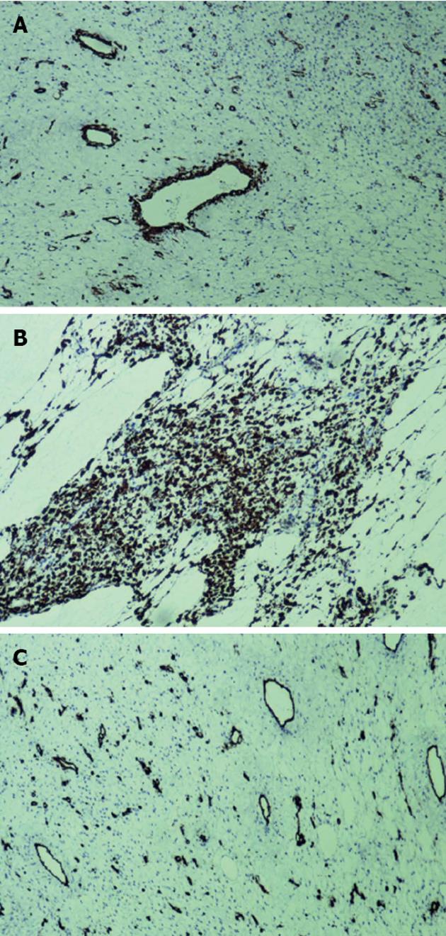
Figure 4 Immunohistochemical analyses.
A: Tumor cells positive to actin (× 10); B: Tumor cells positive to desmin (× 10); C: Tumor cells negative for CD34 (× 10).
- Citation: Stanojević GZ, Mihailović DS, Nestorović MD, Radojković MD, Jovanović MM, Stojanović MP, Branković BB. Case of rectal angioleiomyoma in a female patient. World J Gastroenterol 2013; 19(13): 2114-2117
- URL: https://www.wjgnet.com/1007-9327/full/v19/i13/2114.htm
- DOI: https://dx.doi.org/10.3748/wjg.v19.i13.2114


