Copyright
©2012 Baishideng Publishing Group Co.
World J Gastroenterol. Feb 21, 2012; 18(7): 720-726
Published online Feb 21, 2012. doi: 10.3748/wjg.v18.i7.720
Published online Feb 21, 2012. doi: 10.3748/wjg.v18.i7.720
Figure 1 Ultrasound demonstrated a well-defined, lobulated cystic lesion with fluid debris at the pancreatic head.
The size of the lesion is approximately 5.6 cm × 5.3 cm × 5.7 cm.
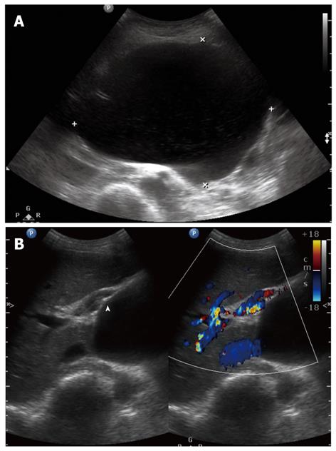
Figure 2 An abdominal ultrasound was performed 4 mo after discharge and revealed an increase in the size of the pancreatic cystic lesion (to 11.
8 cm × 8.7 cm × 11.6 cm) with a clear fluid content (A). Portal vein compression (arrowhead) and varices in the porta hepatis were observed (B).
- Citation: Meesiri S. Pancreatic tuberculosis with acquired immunodeficiency syndrome: A case report and systematic review. World J Gastroenterol 2012; 18(7): 720-726
- URL: https://www.wjgnet.com/1007-9327/full/v18/i7/720.htm
- DOI: https://dx.doi.org/10.3748/wjg.v18.i7.720


