Copyright
©2012 Baishideng Publishing Group Co.
World J Gastroenterol. Feb 21, 2012; 18(7): 712-719
Published online Feb 21, 2012. doi: 10.3748/wjg.v18.i7.712
Published online Feb 21, 2012. doi: 10.3748/wjg.v18.i7.712
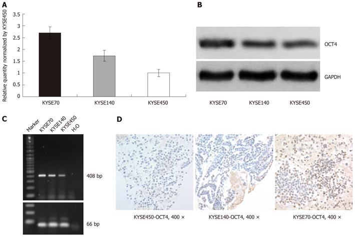
Figure 1 OCT4 expression in esophageal squamous cancer cell lines.
A: The results of real-time polymerase chain reaction (PCR); B: The results of Western blotting; C: The results of conventional PCR; D: The results of immunocytochemistry. GAPDH: Glyceraldehyde-3-phosphate dehydrogenase.
Figure 2 Evaluation of immunohistochemistry staining for OCT4 in tissue array materials of esophageal squamous cell carcinomas (multiplying the extent by intensity, × 400).
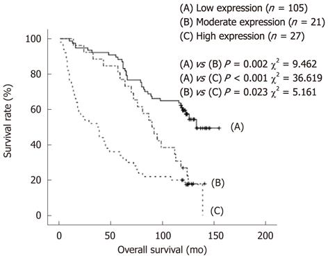
Figure 3 Kaplan-Meier survival curves for esophageal squamous cell carcinoma patients with regard to OCT4 protein expression.
- Citation: He W, Li K, Wang F, Qin YR, Fan QX. Expression of OCT4 in human esophageal squamous cell carcinoma is significantly associated with poorer prognosis. World J Gastroenterol 2012; 18(7): 712-719
- URL: https://www.wjgnet.com/1007-9327/full/v18/i7/712.htm
- DOI: https://dx.doi.org/10.3748/wjg.v18.i7.712


