Copyright
©2012 Baishideng Publishing Group Co.
World J Gastroenterol. Feb 21, 2012; 18(7): 679-684
Published online Feb 21, 2012. doi: 10.3748/wjg.v18.i7.679
Published online Feb 21, 2012. doi: 10.3748/wjg.v18.i7.679
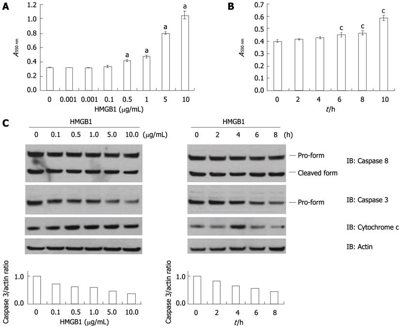
Figure 1 High-mobility group box 1 enhances hepatocyte apoptosis via a mitochondrial pathway.
A: Huh-BAT cells were treated with high-mobility group box 1 (HMGB1) (0, 0.001, 0.01, 0.1, 0.5, 1, 5 and 10 μg/mL) for 6 h. Apoptosis was quantified using an APO Percentage apoptosis assay kit. Data are expressed as the mean ± SD of three individual experiments. aP < 0.05, vs HMGB1 0 μg/mL; B: Huh-BAT cells were treated with 10 μg/mL of HMGB1 for the indicated time periods. cP < 0.05, vs 0 h; C: Huh-BAT cells were treated with HMGB1 (0 μg/mL, 0.1 μg/mL, 0.5 μg/mL, 1, 5 μg/mL and 10 μg/mL) for 6 h (left column), or with 10 μg/mL of HMGB1 for the indicated time periods (right column). Cells were lysed at the indicated time points, and immunoblot analysis was performed using anti-caspase 8 and anti-caspase 3 antibodies. Mitochondrial and cytosolic extracts were also isolated, and equivalent amounts of cytosolic protein were immunoblotted with an anti-cytochrome c antibody. Immunoblot analysis using an anti-actin antibody was performed as a control for protein loading.
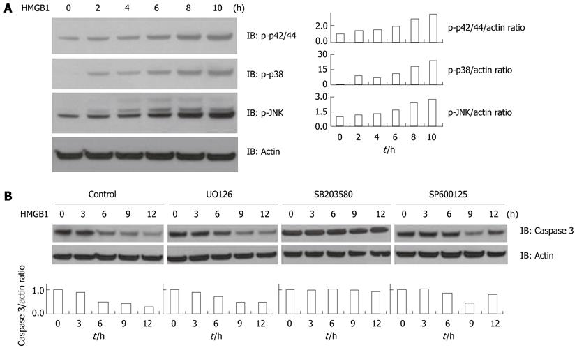
Figure 2 High-mobility group box 1-induced hepatocyte apoptosis is p38-dependent.
A: Huh-BAT cells were treated with 10 μg/mL of high-mobility group box 1 (HMGB1) for the indicated time periods. Cells were lysed at the indicated time points, and immunoblot analysis was performed on cell lysates using antibodies specific for the phosphorylated forms of p42/p44, p38, or c-Jun N-terminal kinase (JNK); B: Huh-BAT cells were pretreated with mitogen activated protein kinase inhibitors U0126 (30 μmol/L), SB203580 (10 μmol/L), or SP600125 (20 μmol/L), or media (control) for 12 h. Cells were then treated with 10 μg/mL of HMGB1. Cells were lysed at the indicated time points, and immunoblot analysis was performed on cell lysates using anti-caspase 3 and anti-actin antibodies.
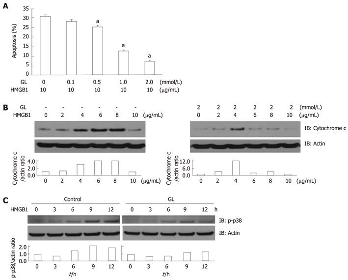
Figure 3 Glycyrrhizin attenuates high-mobility group box 1-induced hepatocyte apoptosis.
A: Huh-BAT cells were pretreated with glycyrrhizin (GL) (0, 0.1, 0.5, 1.0 and 2.0 mmol/L) for 12 h. Cells were then treated with 10 μg/mL of high-mobility group box 1 (HMGB1) for 6 h. Apoptosis was quantified by 4',6-diamidino-2-phenylindole staining and fluorescence microscopy. Data are expressed as the mean ± SD of three individual experiments. aP < 0.05, vs GL 0 mmo/L; B: Huh-BAT cells were pretreated with GL (2.0 mmol/L) for 12 h prior to adding HMGB1 (0-10 μg/mL). Mitochondrial and cytosolic extracts were isolated after 6 h of HMGB1 treatment, and equivalent amounts of cytosolic protein were immunoblotted with an anti-cytochrome c antibody. Immunoblot analysis using an anti-actin antibody was performed as a control for protein loading; C: Huh-BAT cells were pretreated with GL (2.0 mmol/L) for 12 h prior to being incubated with HMGB1. Cells were then lysed at the indicated time points, and immunoblot analysis was performed using antibodies specific for the phosphorylated forms of p38 and actin.
- Citation: Gwak GY, Moon TG, Lee DH, Yoo BC. Glycyrrhizin attenuates HMGB1-induced hepatocyte apoptosis by inhibiting the p38-dependent mitochondrial pathway. World J Gastroenterol 2012; 18(7): 679-684
- URL: https://www.wjgnet.com/1007-9327/full/v18/i7/679.htm
- DOI: https://dx.doi.org/10.3748/wjg.v18.i7.679


