Copyright
©2012 Baishideng Publishing Group Co.
World J Gastroenterol. Feb 21, 2012; 18(7): 646-653
Published online Feb 21, 2012. doi: 10.3748/wjg.v18.i7.646
Published online Feb 21, 2012. doi: 10.3748/wjg.v18.i7.646
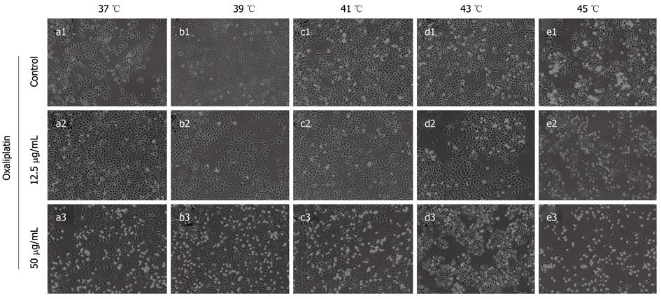
Figure 1 Effect of thermochemotherapy on human colon carcinoma Lovo cells.
Cells were treated with oxaliplatin (12.5 μg/mL and 50 μg/mL) at different temperatures (37 °C, 39 °C, 41 °C, 43 °C or 45 °C) for 1 h and then cultured under normal conditions for 24 h. The cells were observed under inverted phase contrast microscope (Olympus, Japan) after completion of thermal therapy or thermochemotherapy.
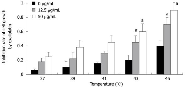
Figure 2 Effect of temperature on the proliferation of Lovo cells by MTT assay.
Lovo cells were treated with oxaliplatin (12.5 μg/mL and 50 μg/mL) at 37 °C, 39 °C, 41 °C, 43 °C or 45 °C. The control group only used thermal therapy but not oxaliplatin. Statistically significant differences were observed between the two groups. Lovo cell viability was determined by MTT assay. The white bars are the results of the oxaliplatin treatment for 1 h at 37 °C, 39 °C, 41 °C, 43 °C, and 45 °C followed by 24 h recovery. The results of three independent experiments are reported as mean ± SD. aP < 0.05 vs 43 °C or 45 °C vs 37 °C.
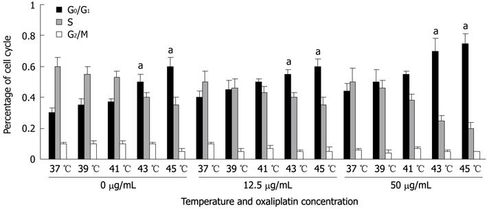
Figure 3 Effect of temperature on cell cycle and apoptosis detected by flow cytometry.
Lovo cells were treated with oxaliplatin (12.5 μg/mL and 50 μg/mL) at 37 °C, 39 °C, 41 °C, 43 °C or 45 °C for 1 h. The different bars represent the percentage of Lovo cells at different phases of the cell cycle at different temperature points. aP < 0.05 vs 43 °C or 45 °C vs 37 °C.
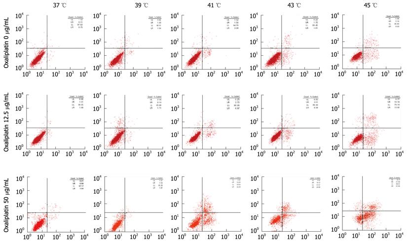
Figure 4 Measurement of Lovo cell apoptosis using apoptosis detection kit.
Data are presented as dot plots in which the vertical axis represents propidium iodide (PI)-positive cells and the horizontal axis annexin V-positive cells. The upper left quadrant region contains necrotic (PI-positive) cells, the upper right region contains the late stage of apoptotic and necrotic (PI- and annexin V-positive) cells, the lower left region contains viable non-apoptotic (PI- and annexin-V-negative) cells, and the lower right region contains early apoptotic (PI-unstained and annexin-V-positive) cells.
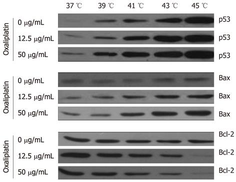
Figure 5 Effect of thermal therapy on p53, Bax and Bcl-2 detected by Western blotting.
Lovo cells were treated with 12.5 μg/mL or 50 μg/mL oxaliplatin for 1 h at 37 °C, 39 °C, 41 °C, 43 °C or 45 °C. Cells were harvested, and total proteins were extracted and immunoblotted for p53, Bax and Bcl-2. The values represent means ± SD of at least three separate experiments. Beta-actin was used as loading control (data not shown).
- Citation: Zhang XL, Hu AB, Cui SZ, Wei HB. Thermotherapy enhances oxaliplatin-induced cytotoxicity in human colon carcinoma cells. World J Gastroenterol 2012; 18(7): 646-653
- URL: https://www.wjgnet.com/1007-9327/full/v18/i7/646.htm
- DOI: https://dx.doi.org/10.3748/wjg.v18.i7.646


